
Presentation
Pain and propstosis in the left eye.
Patient Data
Note: This case has been tagged as "legacy" as it no longer meets image preparation and/or other case publication guidelines.

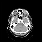


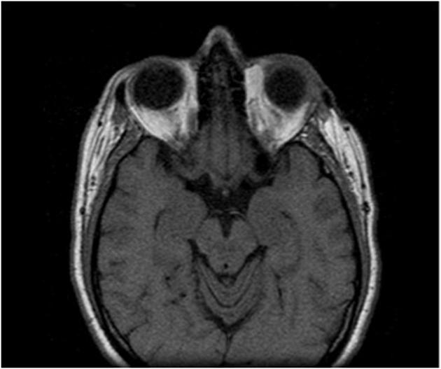
Selected images of CT scan showing a soft tissue hyperattenuating mass in the superior lateral corner of the left orbit which has vivid contrast enhancement.

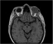
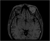
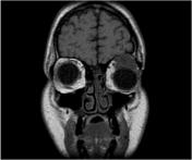

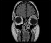
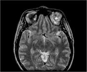
The left orbit anterior mass in the topography of the lacrimal gland is shown with a low T1 signal, a high heterogenous signal on T2 with a small cystic area, and vivid contrast enhancement.
Case Discussion
Adenoid cystic carcinoma is a type of a malignant tumor that affects glandular structures. Orbital adenoid cystic carcinoma usually occurs in patients aged 20-50 years.
This patient had excisional removal of the mass and the final diagnosis was confirmed by histology.


 Unable to process the form. Check for errors and try again.