
Presentation
Follow-up incidental finding.
Patient Data
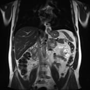

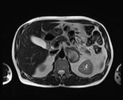
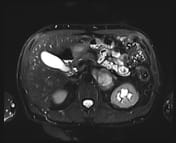
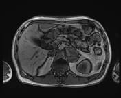
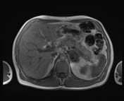
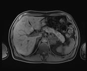
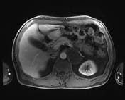
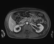
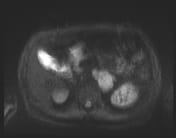
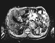
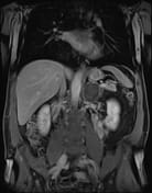
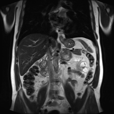
Large left adrenal mass with a low T2 signal rim and high T2 signal center. The mass is lobulated but well marginated. Mild progressive peripheral enhancement. No internal macroscopic fat or microscopic lipid. No strong diffusion restriction.
The patient went on to a left adrenalectomy.
HISTOPATHOLOGY
MACROSCOPIC:
Left adrenal gland – morcellated fragments of the adrenal gland and fat in aggregate 60 x 45 x 20 mm. There is also an intact solid rubbery grey mass 55 x 45 x 40 mm and a separate well-circumscribed rubbery grey nodule 22 x 15 x 7 mm.
The cut surface of both nodules shows a solid slightly whorled grey/white cut surface. No hemorrhage or necrosis is visible macroscopically.
The lesion appears to have been shelled out and margins cannot be assessed. The morcellated fragments of the adrenal gland show no definite mass lesion.
Representative sections from smaller nodules in block 1A, sections from larger nodules in blocks 1B to 1D, and adrenal glands in blocks 1F and 1G.
MICROSCOPIC:
The sections taken from small and large nodule show similar histologic features. These are composed of spindle cell lesions with well circumscribed edge and fascicular/whorled architecture. There are short intersecting fascicles of spindle cells with elongated, wavy nuclei. These spindle cells show focal, mild to moderate variation in nuclear size and shape. No mitotic activity. No necrosis identified. There are randomly distributed scattered ganglion cells.
The surrounding stroma is composed of fibrous and myxoid stroma. No immature, neuroblastic type cells present. Scattered mast cells and occasional lymphoid aggregates are also identified. There is focal calcification (block 1C). Ki–67 shows a proliferative index of less than 1%. S100 immunohistochemistry shows positive staining in the lesional cells.
The completeness of excision cannot be commented upon due to the fragmented nature of the specimen. The features are consistent with ganglioneuroma/s. The background adrenal gland shows no other significant histological abnormality.
DIAGNOSIS:
Left adrenal gland: ganglioneuroma/s.
Case Discussion
Ganglioneuromas are a rare cause of an adrenal mass, and the adrenal gland is a less common site for ganglioneuromas.
There are no specific imaging features for adrenal ganglioneuromas and the imaging differential diagnosis includes 1:
ganglioneuroblastoma
neuroblastoma
composite pheochromocytoma
adrenal cortical adenoma
adrenocortical carcinoma


 Unable to process the form. Check for errors and try again.