Presentation
Abnormal vaginal bleeding
Patient Data


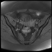

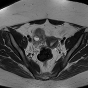

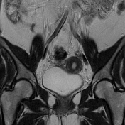

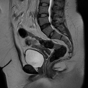

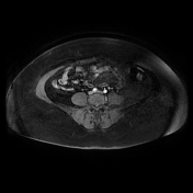

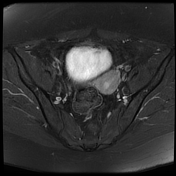

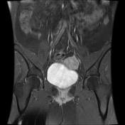

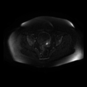

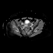

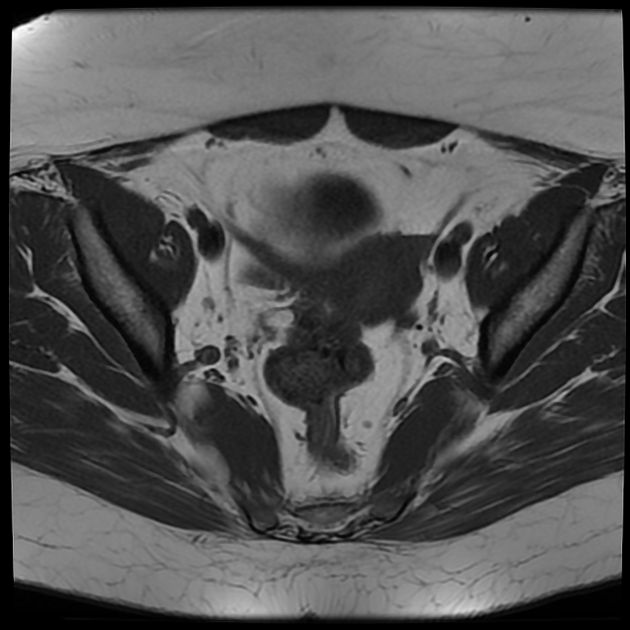
A well-defined lobulated right ovarian mass (5.5 x 5 x 4.5 cm) of intermediate signal intensity on T1WI, intermediate to high signal intensity on T2WI, containing areas of high signal on T1WI and T2WI with hemosiderin sediment on T2WI (hemorrhage within locules). The solid component shows moderate and heterogeneous enhancement following IV contrast. The DWI / ADC show restricted diffusion.
Left ovary and uterus appear normal with no significant endometrial thickening (thickness = 7 mm)
No pelvic lymphadenopathy or effusion is seen.
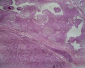
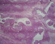
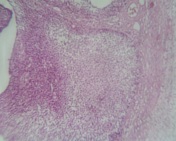
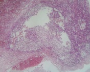
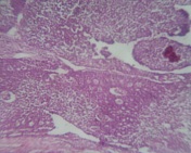
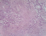
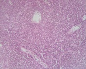

Macroscopically: the tumor was well-encapsulated, measuring 5 x 5.2 cm, with a smooth outer surface, heterogeneous in appearance: whitish solid with cystic components containing hemorrhage.
Microscopically: it appears as a well-defined, encapsulated, tumoral proliferation composed of cell layers, multinodular structures, trabecular patterns and some follicular territories of variable size delineating fibro-hyaline structures corresponding to the "Call-Exner bodies"
Immunohistochemistry:
-vimentin: positive.
-CK7 and CK20: shows negativity of the proliferation
-actin: positive
-AE 1 / AE 3 shows a positive marking (focally)
-EMA: negative
-Inhibin: positive, calretinin: positive
Case Discussion
The patient had right salpingo-oophorectomy with pathologically proven adult granulosa cell tumor
It is considered as the most frequent subtype of granulosa cell tumors of the ovary (95%) and is commoner than the juvenile form.
Additional contributor: Selma Nezar MD, anatomopathologist, Batna, Algeria


 Unable to process the form. Check for errors and try again.