Patient Data
Age: 60 years
Gender: Male
From the case:
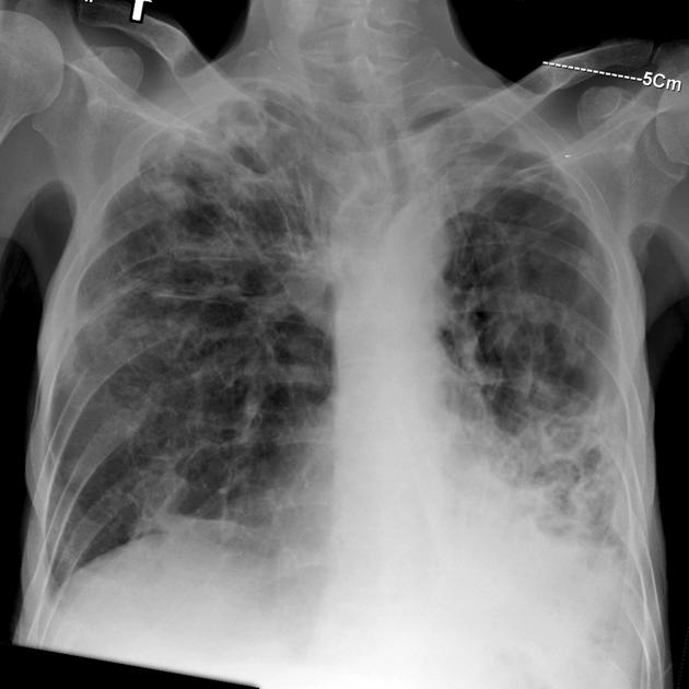
Advanced pulmonary tuberculosis
Download
Info

CXR demonstrates signs of advanced pulmonary TB: Bilateral, coarse, predominantly upper lobe fibrosis with pleural thickening and callus formation, large, confluous cavities, diffuse consolidated areas. Note the resultant abnormally elevated position of the hila, better appreciated on the right.
From the case:
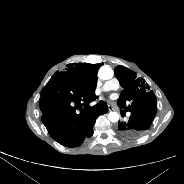
Advanced pulmonary tuberculosis


Download
Info

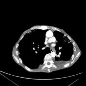
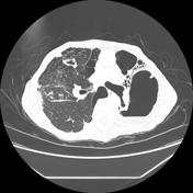
CT demonstrates the same findings as the plain film, in addition basal traction bronchiectasis and severe honeycombing can be discerned corresponding to the consolidated areas on CXR. Gas-fluid level can also be observed in the left lower lobe cavities.
Case Discussion
Advanced pulmonary TB.


 Unable to process the form. Check for errors and try again.