
Presentation
Neurologically abnormal with an enlarged head.
Patient Data
Age: Neonate
Gender: Male


Download
Info

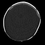
Non-contrast CT demonstrates a thin rim of cerebral tissue anteriorly, without midline cleft. There is a large CSF-density space posteriorly. There is no evidence of a falx cerebri. The cerebellum is malformed with abnormal appearance of the fourth ventricle. Brainstem is visualized.
In addition there are a number of other midline abnormalities:
- bony defect in the occipital bone with an occipital meningiocoele
- cleft lip and palate
- under-developed nose and hypotelorism
Case Discussion
This case demonstrates the typical appearances of alobar holoprosencephaly, incompatible with long term survival.


 Unable to process the form. Check for errors and try again.