Presentation
Neck pain and bilateral upper limb numbness for two years.
Patient Data

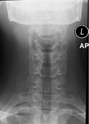
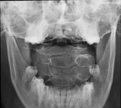
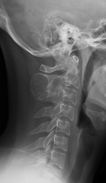
Well-defined expansile radiolucent lesion in the spinous process of the 2nd cervical vertebra. No fracture or dislocation is seen.
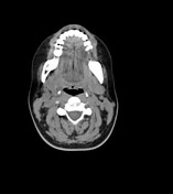

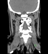

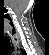

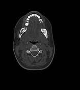

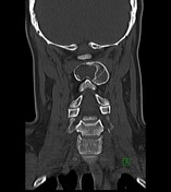

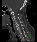

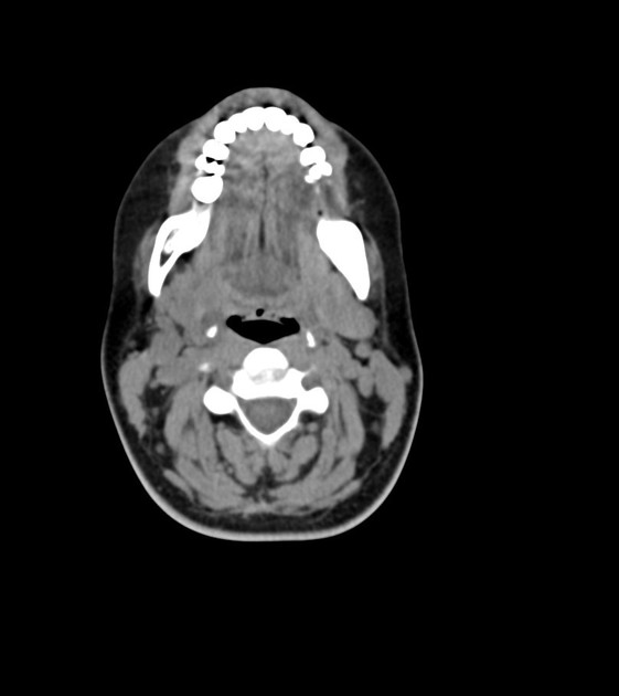
An expansile radiolucent lesion is seen in the spinous process of C2 vertebra, with mild extension into laminae (left more than the right). No fluid-fluid or blood-fluid levels are seen in it. No cortical disruption or associated soft tissue component is noted. No intraspinal extension is noted.
Incidental partially depicted azygos fissure.
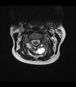

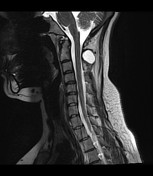

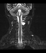

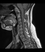

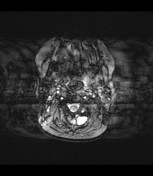

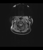

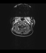

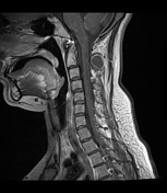

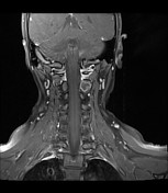

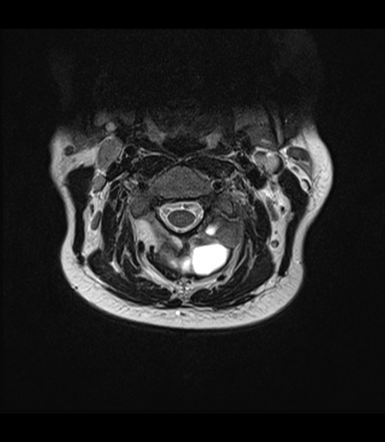
Mildly expansile multilocular cystic lesion, involving the spinous process and laminae of the C2 vertebra. No fluid-fluid levels or hemorrhage is noted in it. It has peripheral enhancement on the post-contrast study; however, has no enhancing internal solid component. No intraspinal extension is noted.
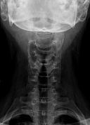
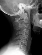
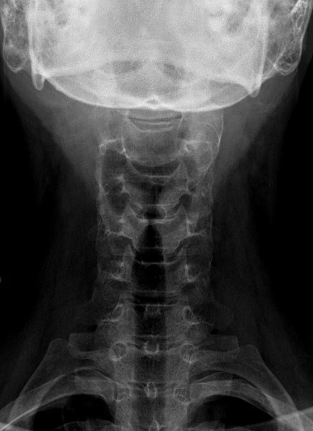
Large expansile cystic lesion involving the spinous process of C2 vertebra, showing no gross interval change.


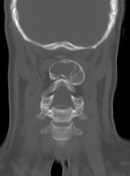

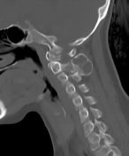

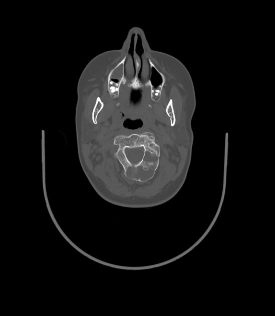
No significant interval change is seen in the expansile bony lesion involving the posterior elements of the C2 vertebra.
Pre-operative diagnosis: Well-defined expansile radiolucent lesion involving the posterior elements of the 2nd cervical vertebra, showing no significant interval change on the follow-up images, suggestive of a benign bony tumor, likely aneurysmal bone cyst.
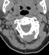

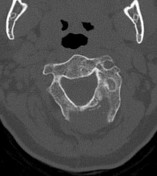

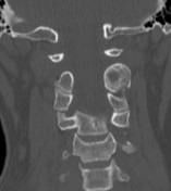

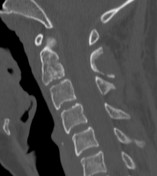

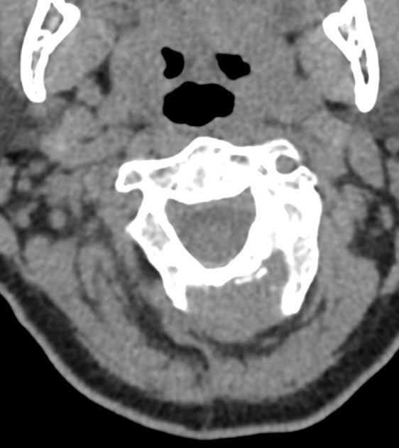
Evidence of surgical intervention (aspiration of cystic contents/curettage and bone grafting) is noted in the C2 posterior arch tumor.
Case Discussion
Procedure: Debulking of the shell, aspiration of the contents, microscopic resection of cystic components with preservation of the inner shell and instillation of artificial bone for bone remodeling and healing.
Gross description: The specimens received in two containers:, labeled with the patient's name and medical record number. Part 1 (received fresh for intraoperative consultation), labeled "spinal tumor” consists of multiple fragments of bony and soft hemorrhagic tissue measuring in aggregate 2.0 x 2.0 cm. Part 2 (received in formalin), labeled "spinal tumor" consists of multiple fragments of bony and soft hemorrhagic tissue measuring 3.0 x 2.0 x 0.4 cm.
Microscopy: Sections show blood, reactive trabecular bone and a fragment of fibrous tissue (septum) which forms a cystic space that is filled with blood. Parallel arrays of osteoid trabeculae along the septum and rare osteoclast-like giant cells are also noted. Negative for malignancy.
Diagnosis: Aneurysmal bone cyst.


 Unable to process the form. Check for errors and try again.