
Presentation
A child with respiratory distress after elective surgery for a tracheocutaneous fistula.
Patient Data
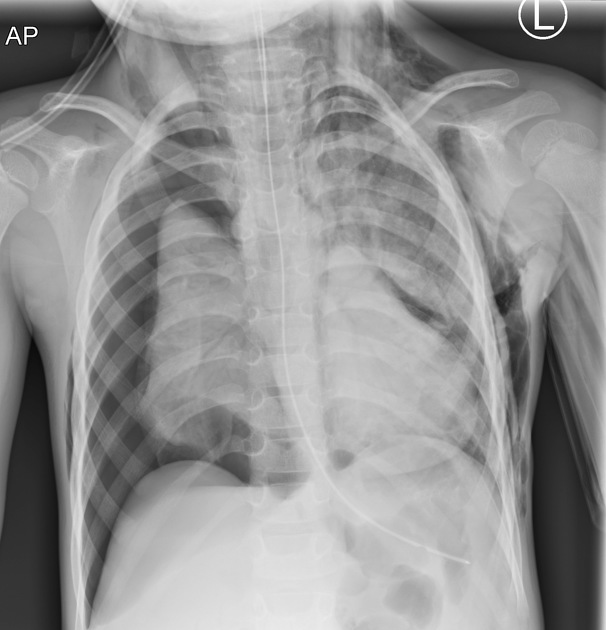
Frontal radiograph showing the most life-threatening finding of right-sided tension pneumothorax with left-sided mediastinal shift; this should be immediately decompressed. Extensive left-sided subcutaneous emphysema is also featured. However, another pertinent finding is of a well-defined structure over the left hemithorax midzone which appears to be 'lifted' off the left heart border. This is the angel wing or spinnaker sign representing the pediatric thymus in pneumomediastinum.
Nasoenteric tube is correctly placed in the stomach.
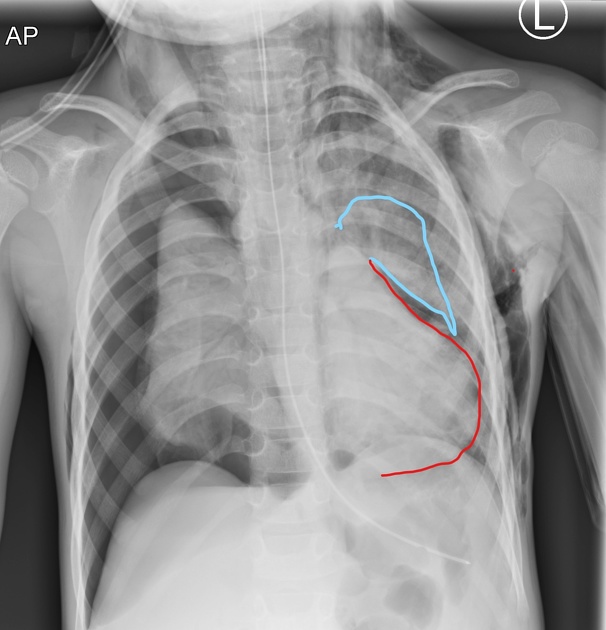
Same frontal radiograph highlighting the angel wing thymus (blue lining) lifted off the left heart border (red lining)
Case Discussion
Certain plain radiographic signs of pediatric pneumomediastinum are unique compared to adults:
- air can lift the relatively large thymus gland off from the heart border to give the angel wing or spinnaker sign
- another sign is air inferior to the mediastinum/superior to the diaphragmatic central tendon which can give the heart a conical appearance described as the Haystack sign after Monet's painting of the same name
These signs are important to be aware of in the young patient as pediatric pneumomediastinum can be otherwise occult.
This case represents an iatrogenic etiology but a careful review of the case is usually required when there is no history of surgical intervention to establish a differential (e.g. infection, esophageal perforation, etc).
The spinnaker or angel wing sign should also not be confused with the thymic sail sign which refers to the pointy/triangular inferior edge of the thymus in normal cases.


 Unable to process the form. Check for errors and try again.