
Presentation
Pulsatile upper abdominal mass on physical exam.
Patient Data
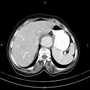

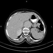

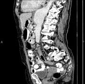
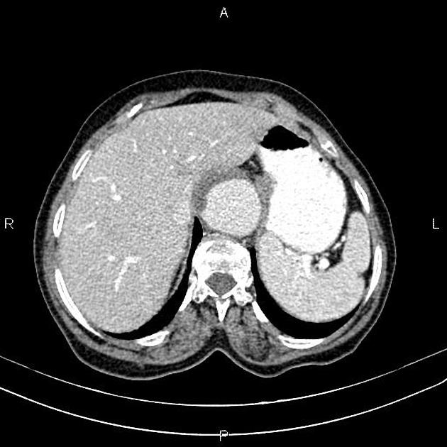
Fusiform aneurysmal dilatation of the lower thoracic and upper abdominal aorta is present, measuring 5 cm in maximum diameter and about 8 cm in length. Mild mural thrombosis is observed at the right anterolateral aspect.
Intra and extrahepatic bile ducts are mildly dilated, and CBD measured 10 mm in caliber.
A 30 mm diverticulum is evident at the medial aspect of the second portion of the duodenum that seems to be compressed distal of CBD, suggesting Lemmel syndrome.
A few non-enhanced simple cortical cysts are seen in both kidneys.
Degenerative changes such as osteophytosis are seen in the lumbar spine.
Grade I spondylolisthesis of L5 on S1 is present.
Case Discussion
Open repair of the aortic aneurysm was performed with a vascular graft at a referral center. The patient is doing well postoperatively.


 Unable to process the form. Check for errors and try again.