
Presentation
Several year history of small bowel Crohn disease. Recently presented with diarrhea.
Patient Data
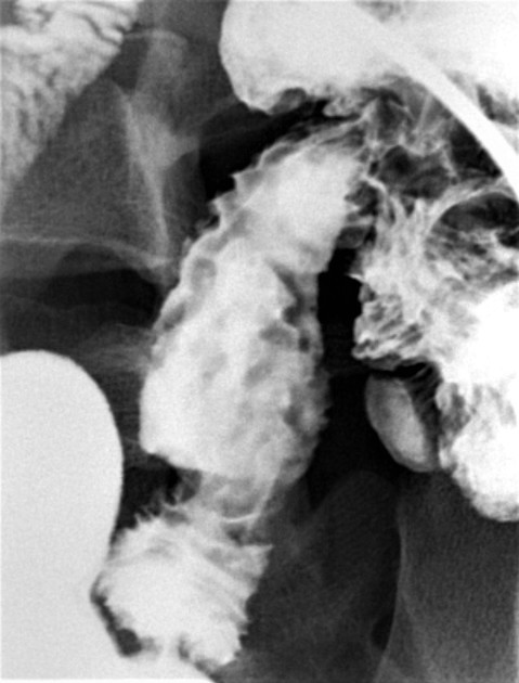
There are mildly thickened nodular folds in the terminal ileum, compatible with inflammation. Superimposed on these are tiny oval lucencies with faint central dots of barium, compatible with mucosal ulcerations (aphthous ulcerations).

Arrows point to the small oval lucencies with central "umbilicated" dots of barium, compatible with small aphthous ulcers.
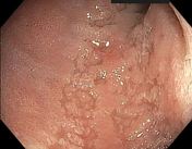
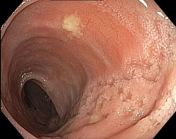
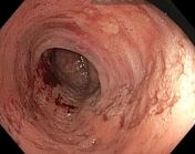

Images of the intubated terminal ileum during colonoscopy. There is scarring and ulceration in the terminal ileum compatible with inflammation from Crohn disease. Aphthous ulcers are superimposed on more chronic-appearing linear ulceration/scarring.
Case Discussion
Aphthous ulcers often appear as radiolucent ovals ("halos") with central punctate dots of barium These represent surface mucosal erosions (on the surface of hyperplastic lymphoid follicles) Aphthous ulcers are usually distinct from nearby smooth normal mucosa but they can be clustered and are sometimes found at the edge of more advanced disease
As Crohn disease progresses, individual aphthous ulcers may merge to form larger ulcerations


 Unable to process the form. Check for errors and try again.