
Presentation
Initial symptoms included a holocranial headache which was pulsatile in nature, and accompanied by nausea and vomiting.
Patient Data


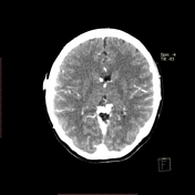
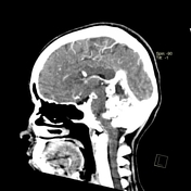
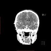
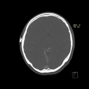
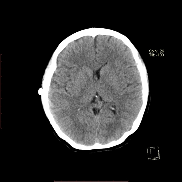
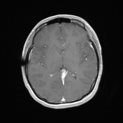
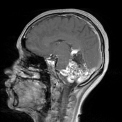
CT shows an arterio-venous malformation in the cerebellar region and a ventriculoperitoneal catheter terminating in the right lateral ventricle.


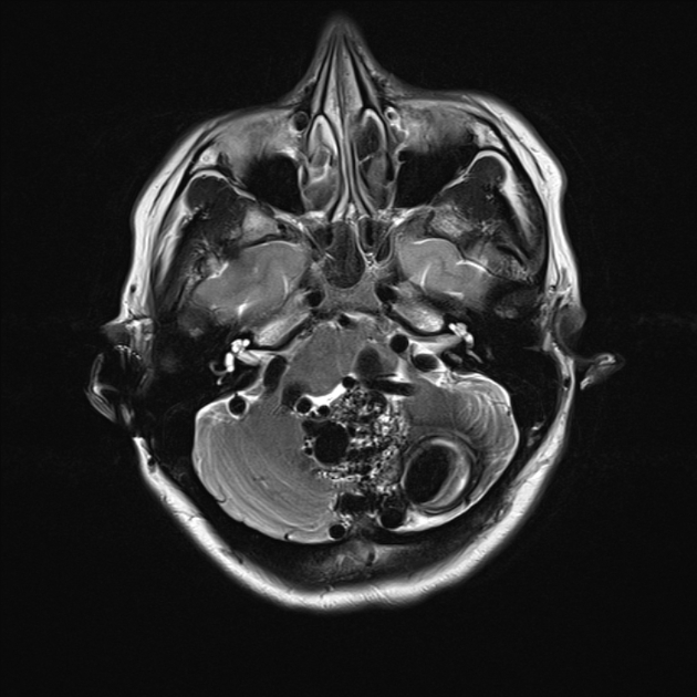
Left cerebellous hemisphere in the qudtrade lobulillo and extension towards vermis.
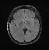
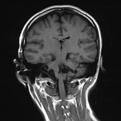
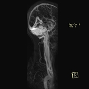
MRI shows multiple images of serpiginous tract, confluent, which present flow voids in T2 with opacification during the arterial phase, with dependent arterial supply from posterior cerebral arteries and venous drainage to the transverse sinus, observing veins with aneurysmatic dilatation in the left cerebellar hemisphere, as a whole it measures 5.7 x 4.1 x 5.6 cm in the rostrodorsal, dorsoventral and lateral direction. This AVM conditions obliteration of the fourth ventricle and anterior displacement of the pons and medulla with compression of the left cerebellar peduncle as well as rectification of the tentorium.
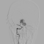
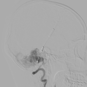
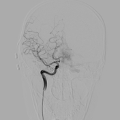
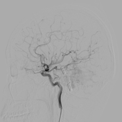
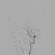
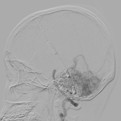

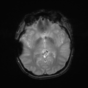
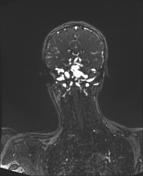
During the selective opacification of the left vertebral artery, in the early arterial phase of the study, multiple vessels of disorganized appearance that make up a plexiform network are identified, dependent on the right posterior cerebral artery as well as the ipsilateral posteroinferior cerebellar artery, which converge and nourish a niche vascular visualized mainly during the late arterial and capillary phase; observing consecutive and early opacification of the outlet vein with bilateral drainage to the transverse sinus, in addition to observing aneurysmal dilation of the draining veins during the venous phases of the study. These findings are in relation to arteriovenous malformation.
Case Discussion
This patient has a single arteriovenous malformation located in the posterior fossa that mainly affects the cerebellum with compression of the fourth ventricle. According to the Spetzler-Martin arteriovenous malformation (AVM) grading system, this is classified as grade IV, with a nidus size greater than 6 cm, located in eloquent brain, with feeding branches of the posterior cerebral artery and posteroinferior cerebellar artery and drainage to the transverse sinus.


 Unable to process the form. Check for errors and try again.