
Presentation
Left sided pulsatile tinnitus.
Patient Data
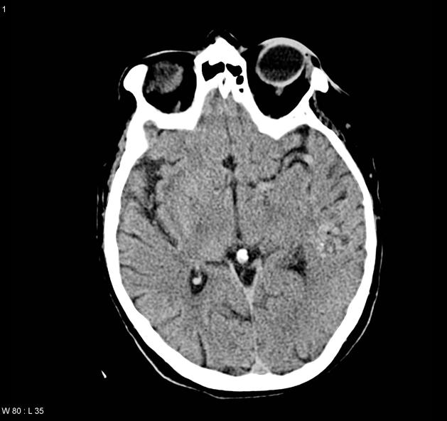
Non contrast CTB demonstrates a region of irregular density in the left temporal lobe, slightly hyperdense to adjacent white matter. A few flecks of calcification are seen within it.
The left internal carotid artery (ICA) and middle cerebral branches appear enlarged, and there is the suggestion of enlarged tubular structures over the surface of the temporal lobe.

MIP CTA confirms the presence of a left temporal lobe arteriovenous malformation (AVM) which appears to receive the vast majority of its supply from the left middle cerebral artery. Contribution from the posterior cerebral artery is although likely via the lateral posterior choroidal arteries. Contribution from anterior cerebral artery and meningeal arteries cannot be excluded.
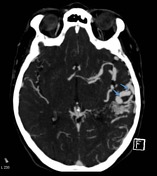
Drainage appears to be predominantly superficial, over the temporal lobe and into the transverse / sigmoid sinus. The superior petrosal sinus appears plump and likely also provides some drainage.
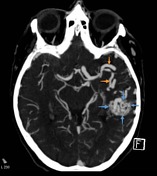
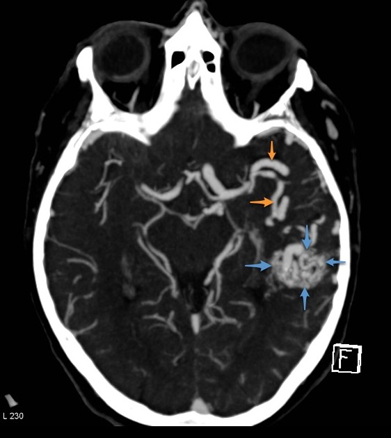
- Fig 1: nidus (blue arrows) and enlarged middle cerebral arteries (orange arrows).
- Fig 2: draining veins (blue arrows)
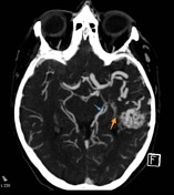
- Fig 3: enlarged posterior cerebral artery (blue arrow) with likely contribution via the lateral posterior choroidal arteries. Small branches can be seen extending from the choroid plexus to the nidus (orange arrow).
Case Discussion
This case demonstrates characteristic CT and CTA appearances of a cerebral arteriovenous malformation (AVM). Given its location in the dominant (assuming right handed patient) temporal lobe, likely deep drainage and a small (<3cm) nidus, this would have a S-M grade of 3. Catheter angiography is required to fully assess, and potentially treat, this lesion.


 Unable to process the form. Check for errors and try again.