
Presentation
Palpable midline parietal soft tissue mass since birth.
Patient Data


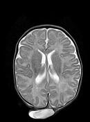


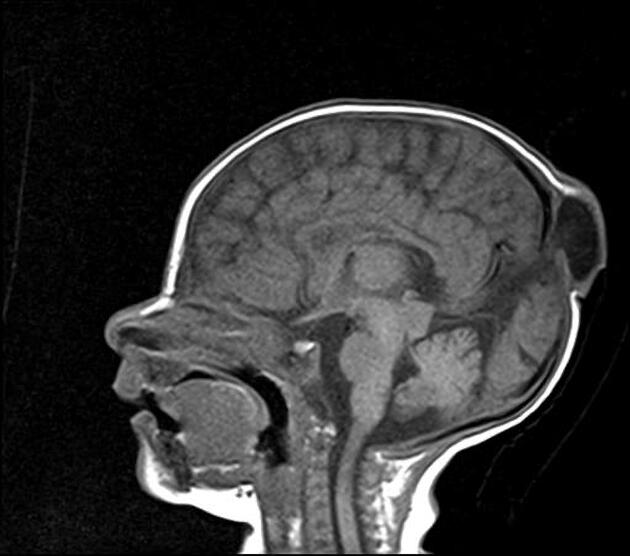
Midline subgaleal parietal cystic mass at the posterior fontanelle with a fibrous stalk connecting it to the dura via a calvarial defect. It displays a CSF signal on all sequences with some internal linear structures representing probably meninges with glial and neural rests.
Persistent primitive falcine vein vertically oriented to the subgaleal scalp mass with a high position of the tentorium cerebelli and prominent superior cerebellar cistern.
The straight sinus is absent as well as the vein of Galen.
Case Discussion
The MRI features are most consistent with an atretic parietal cephalocele.
Atretic parietal cephaloceles are thought to represent involuted true cephalocele (meningocele or encephalocele) connected to the dura mater via a fibrous stalk.


 Unable to process the form. Check for errors and try again.