
Presentation
Fever and cough for 2 days. Shortness of breath for one day.
Patient Data


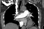
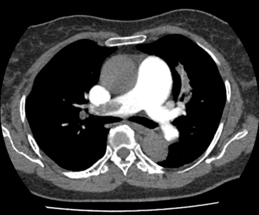
Massive filling defects seen in bilateral distal main pulmonary arteries, lobar arteries, segmental and subsegmental branches. Almost all branches show no contrast opacifcation.
The main pulmonary trunk and bilateral main pulmonary arteries are dilated.
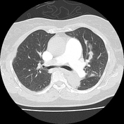
Lung window: collapse/consolidation and patchy peripheral wedge shaped consolidation at the apicoposterior segment of left upper lobe suggestive of pulmonary infarcts.
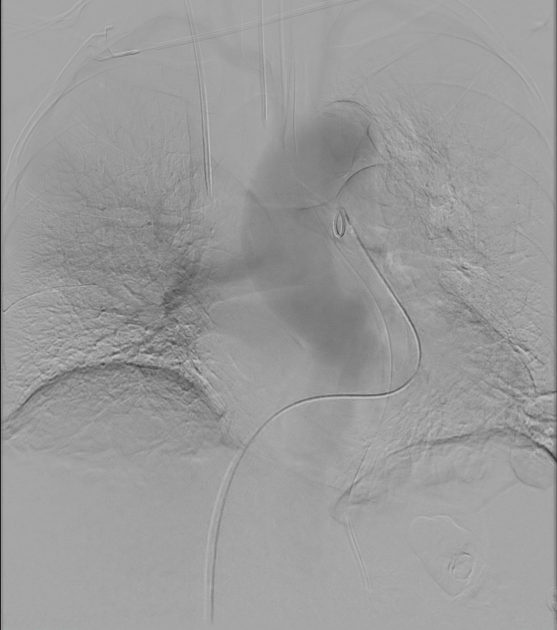
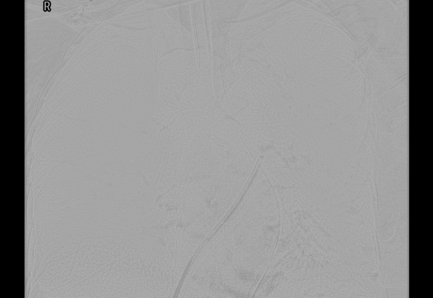
Improved flow noted at the right superior pulmonary artery. However, there is still residual filling defect suggestive of chronic thrombus.
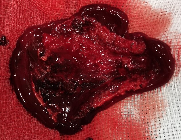
Blood clots aspirated after vacuum assisted thrombectomy.
Case Discussion
The patient was shown to have massive bilateral pulmonary embolism on CT pulmonary angiogram. An emergency vacuum-assisted thrombectomy was done. However only about 30-40% of the blood clots were aspirated. The remaining 60-70% of the blood clots remained in the pulmonary vessels. The patient remained in the ICU after the thrombectomy and passed away a few days later.


 Unable to process the form. Check for errors and try again.