
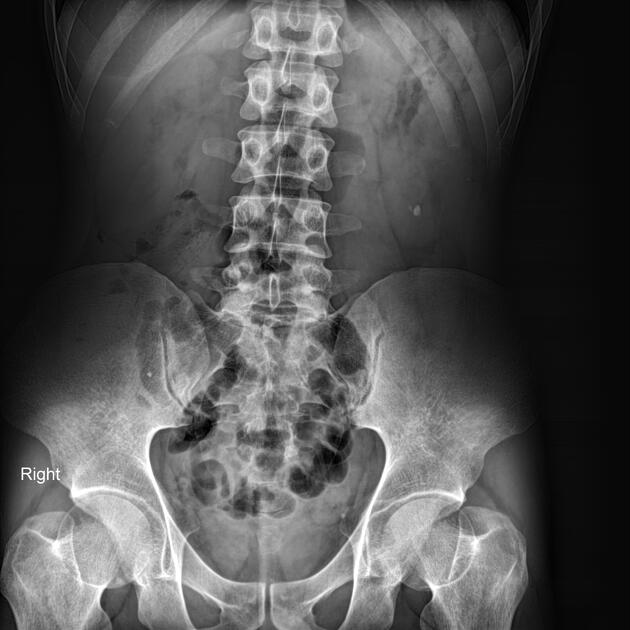
Presentation
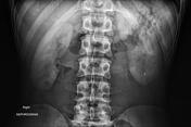
Work up for microscopic hematuria.
Patient Data
Age: 25 years
Gender: Male
From the case:
Bilateral ureteropelvic junction obstruction


Download
Info
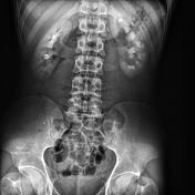
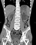
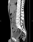
In non-contrast images, a small stone is noted in the lower part of the left renal lodge.
After contrast media administration, the enhancement of both kidneys is non-uniform. In the secretory phase, moderate hydronephrosis without hydroureter is evident bilaterally; the left is more severe than the right causing delayed contrast excretion.
From the case:
Bilateral ureteropelvic junction obstruction


Download
Info
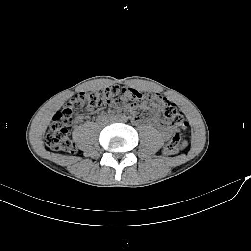
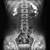
Moderate dilatation of the pelvicalyceal system is seen on both sides without ureter dilatation or ureteral stone.
A stone is also noted in the left kidney's lower part.
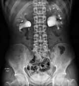
Case Discussion
Typical IVU and CT scan appearance of the bilateral ureteropelvic junction obstruction (UPJO).


 Unable to process the form. Check for errors and try again.