
Presentation
Dyspnea, cough and facial swelling
Patient Data


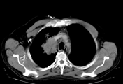
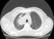
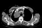
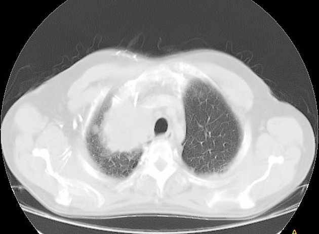
Right upper lobe mass infiltrating the right brachiocephalic vein and superior part of SVC with development of posterior and anterior chest wall collaterals.
Right diaphragmatic paralysis likely due to phrenic nerve infiltration.
Right paratracheal lymphadenopathy.
Diagnosis: T4N1Mx
Case Discussion
most common malignancy in males
age : > 50 years old
sex : M>F
TNM classification:
-
T1
T1a (< 2 cm)
T1b (2-3 cm) surrounded by lung or visceral pleura, without invasion more proximal than lobar bronchus
-
T2
-
T2a (3-5 cm), T2b (5-7 cm), or tumor with any of the following features:
involves main bronchus > 2 cm distal to carina
invades visceral pleura
associated with atelectasis or obstructive pneumonitis that extends to the hilar region but does not involve the entire lung
-
-
T3
tumor > 7 cm or any of the following
directly invades any of the following: chest wall, diaphragm, phrenic nerve, mediastinal pleura, parietal pericardium, main bronchus < 2 cm from carina without involvement of the carina
atelectasis or obstructive pneumonitis of the entire lung
separate tumor nodules in the same lobe
-
T4
tumor of any size that invades the mediastinum, heart, great vessels, trachea, recurrent laryngeal nerve, esophagus, vertebral body, carina, or with separate tumor nodules in ipsilateral lobe.


 Unable to process the form. Check for errors and try again.