
Presentation
Presenting to ED for chest pain no significant past medical history.
Patient Data

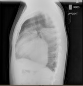
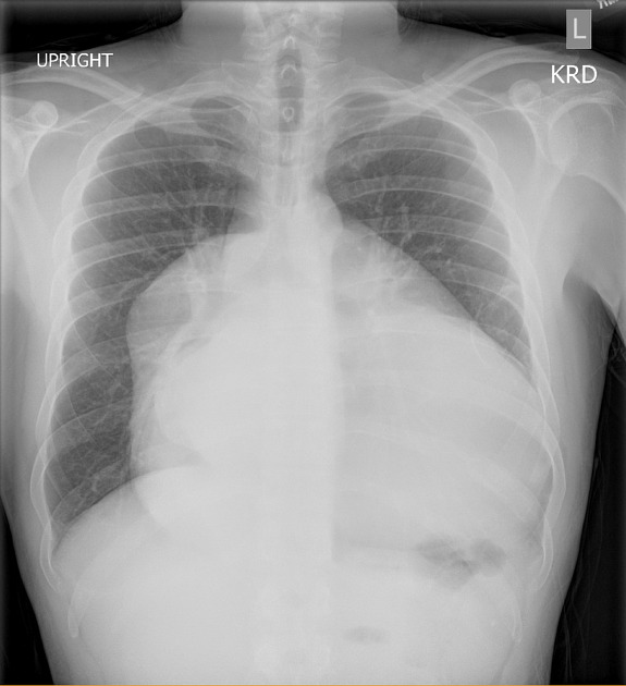
No acute pulmonary findings.
Severe expansion of cardiac silhouette. Unclear etiology consider MRI for further evaluation
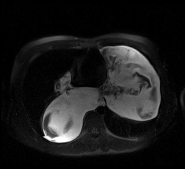
Very large multiloculated cystic lesion in all compartments of the mediastinum creating mass effect and encasement of the trachea and surrounding mediastinal structures. It extends from above the great vessels to the diaphragm. It displays T2 hyperintensity with solid features with loculations. It measures 9 x 11 x 19 cm in the right hemithorax, and 11 x 14 x 12 cm in the left hemithorax. Findings are suggestive of a large bronchogenic cyst.
Case Discussion
This case presents an impressive chest x ray which at first can be very alarming. Further evaluation using other imaging modalities showed a mediastinal mass which turned out to be a bronchogenic cyst. Bronchogenic cysts are relatively benign masses which come from bronchopulmonary foregut dysplasia during development. They are not malignant but can grow to be quite large as seen in this patient and cause mass effect and encasement of surrounding mediastinal structures. This was the case here and the patient was to be followed up for outpatient cardiothoracic surgery evaluation for removal.
The case also brings about the classification and description of the compartments of common mediastinal masses. Anterior mediastinum hosting thymomas, thyroid neoplasms, lymphomas, germ cell tumors. Middle mediastinum hosting most bronchogenic cysts. Posterior mediastinum being host to neurogenic tumors such as schwanommas in the chest.


 Unable to process the form. Check for errors and try again.