
Note: This case has been tagged as "legacy" as it no longer meets image preparation and/or other case publication guidelines.
Download
Info
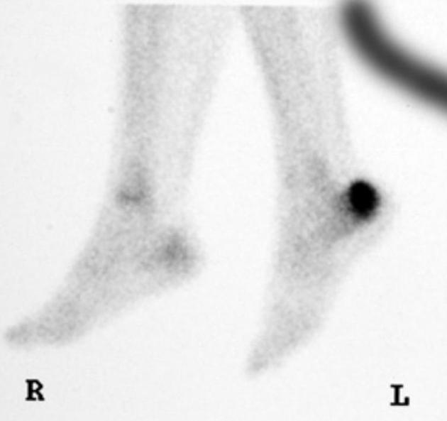
Intense increased uptake in the left calcaneum.
From the case:
Calcaneal stress fracture
Download
Info
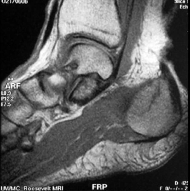
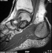
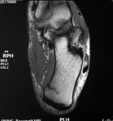
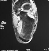
Linear T1 intramedullary signal in the calcaneus with surrounding edema.
Case Discussion
Bone scan and MRI reveal a calcaneal stress fracture.
These images are from Dr. John Hunter's amazing MSK collection. Dr. John Hunter is a professor in the department of radiology (musculoskeletal section) at UC Davis School of Medicine.
This case was donated to Radiopaedia.org by Radswiki.net.


 Unable to process the form. Check for errors and try again.