Presentation
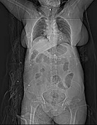
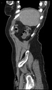
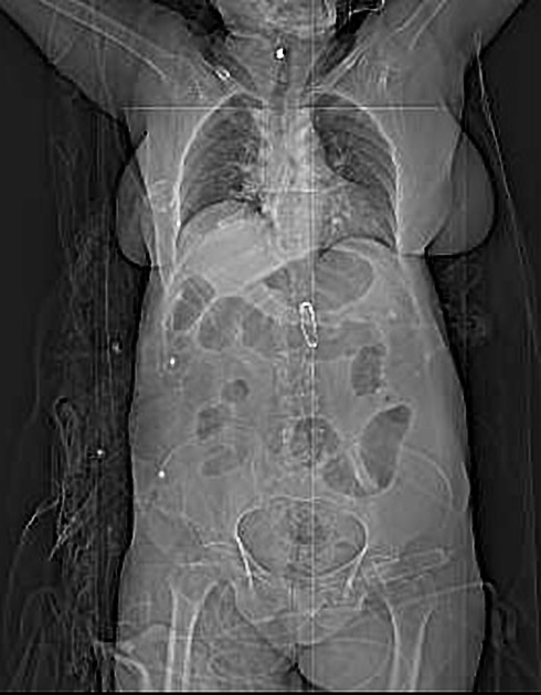
Road traffic accident, in which she sustained a right leg fracture. Incidental finding of a calcified mass in the right breast on CT.
Patient Data
Age: 70 years
Gender: Female
From the case:
Calcified breast lesion (CT)


Download
Info

A hypodense large round mass of fat density with coarse peripheral ring-like wall calcification in the right breast.
Case Discussion
The pattern of calcification in this lesion is benign.
Eggshell calcifications are benign peripheral rim-like calcifications and are typically secondary to fat necrosis or calcification of oil cysts. Typically, they measure <1 mm.
Involuting fibroadenoma can be also another possibility.


 Unable to process the form. Check for errors and try again.