
Presentation
Neglected right hemiparesis. No history of cervical trauma
Patient Data
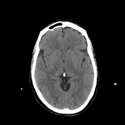

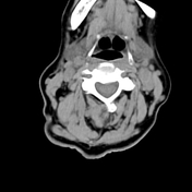

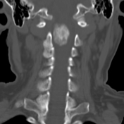
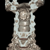
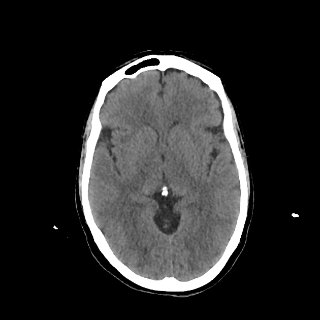
There is a calcified mass within the cervical canal at C2 level below the foramen magnum of posterior location, compressing and displacing the spinal cord anteriorly.

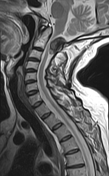
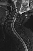
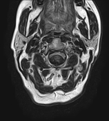
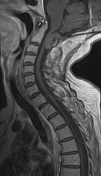
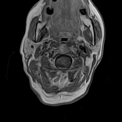
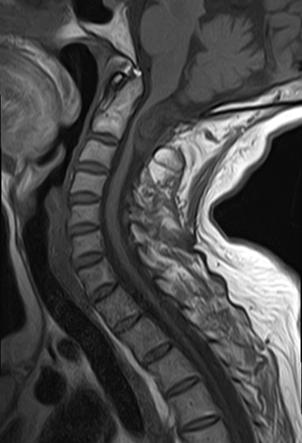
This mass of intradural extramedullary location displays a low signal intensity on T1WI, T2WI, and STIR with posterior broad-based attachment. The postcontrast sequences show a heterogeneous enhancement. The adjacent segment of the spinal cord is compressed and displaced anteriorly with an area of high signal on T2WI/STIR, indicating compressive myelopathy.
Case Discussion
MRI features are most consistent with calcified meningioma of the cervical canal compressing the spinal cord with signs of compressive myelopathy.
On imaging, the differential diagnosis should include a calcified hematoma, but the absence of a history of cervical trauma and the enhancement on postcontrast sequences are the key to the correct diagnosis.
Additional contributors: C. Boukaaba, MD, Z.E Boudiaf, MD


 Unable to process the form. Check for errors and try again.