
Presentation
Chest pain, shortness of breath for 2 weeks. History of asbestos exposure.
Patient Data
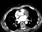

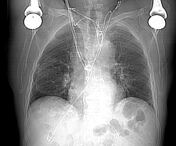
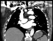
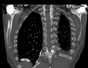
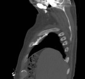

Multiple calcified pleural plaques bilaterally. No evidence of pleural effusion.
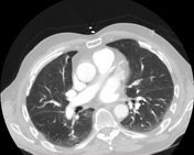
Focal atelectasis in the right upper and both lower lobes.
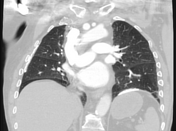
No CT evidence of pulmonary thromboembolism.
The thoracic aorta demonstrates mild atheromatous calcification without evidence of aneurysm.
Coronary artery calcification is noted. There is no pericardial effusion.
Small mediastinal lymph nodes
Anterior wedging and decreased height of T12 vertebral body. Bilateral shoulder arthroplasty.
Case Discussion
Changes characteristic of asbestos related pleural disease with multiple bilateral calcified pleural plaques. These have anterolateral, posteromedial and diaphragmatic predominance but spare the costophrenic angles.
There is dependent atelectasis in both lungs and no evidence of fibrosis (asbestosis).


 Unable to process the form. Check for errors and try again.