
Cardiomediastinal outlines on chest x-ray
Citation, DOI, disclosures and case data
At the time the case was submitted for publication Frank Gaillard had no recorded disclosures.
View Frank Gaillard's current disclosures
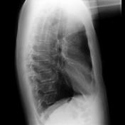

Chest x-ray demonstrate normal cardiomediastinal outlines. No pulmonary or pleural mass identified. There is a minor degree of hyperinflation, which may represent a degree of underlying COPD.
5 case questions available
Q: What makes up (from top to bottom) the right cardiomediastinal border? show answer
A: Right paratracheal stripe, arch of the azygous vein, superior vena cava (or ascending aorta in older pts), right atrium, inferior vena cava (IVC)
Q: What makes up (from top to bottom) the left cardiomediastinal border? show answer
A: Left paratracheal stripe (made up of left common carotid artery, left subclavian artery and the left jugular vein), aortic arch, pulmonary artery, auricle of left atrium, left ventricle.
Q: When visible, where is the aortic nipple and what structure accounts for it? show answer
A: The aortic nipple is a small protuberance from the aortic knuckle, seen on frontal chest radiograph. It is due to the superior intercostal vein.
Q: What makes up (from top to bottom) the anterior cardiomediastinal border? show answer
A: Superior mediastinum (great vessels & thymus), ascending aorta, right ventricular outflow tract, right ventricle.
Q: What makes up (from top to bottom) the posterior cardiomediastinal border? show answer
A: Left atrium and pulmonary veins, left ventricle, inferior vena cava.


Annotated frontal and lateral chest x-ray with structures that account for the mediastinal outline labeled.
Case Discussion
A thorough understanding of the structures which normally contribute to cardiomediastinal outline is essential in being able to interpret chest x-rays and localize abnormalities.
8 articles feature images from this case
220 playlists include this case
Public playlists
- Intern Education - Chest by Wayland Wang ◉ ◈
- UQMS pre-elective CXR tute by Kelvin K Y Ho
- Chest by Magdiel Bishop
- UQMS pre-elective CXR tute by Kelvin K Y Ho
- Normal CXRs by Chris Yu
- 661-Chest and Abdomen Anatomy by Lauren Tollefson
- medical student CXR by Ghadir H Kassab
- ALL by Andrew Mironenkov
- Anaesthetics 2023 by Jarrah Spencer
- Normal Anatomy by Carlos Gray
- 1.1.3 E1 by Elisabeth Ranharter ◉
- CVD congenital by Chandra
- Intern Radiology - Chest X-ray by Craig Hacking ◉
- A&B - Thorax by Jan Philipp Hering
- Intern Radiology - CT chest by Craig Hacking ◉
- Tórax by Luis Miguel
- Mediastinum & Pericardium PART 1 by Varun Tyagi
- Anatomy by Mariangela Alvarado Molinaro
- General 3B by Lisa Guion
- Chest Radiograph by Ian Crosbie
- CXR Q by Dr Ali Basim
- Mediastinum & Pericardium PART 1 by Varun Tyagi
- Chest Xray-abcde normal by Md. Shahriar Anzum Shuvo
- Chest x-rays (iOS pack) by Frank Gaillard ◉ ◈
- Anatomy by Ahmed Reda Bassiouny
- CXR 101: A Primer by Aaron Wong
- Chest by Ashley Hook
- chest by Robert Ligetfalvi
- Exam by Dave Boha
- anatomija_krutine by Tomas Jurevicius
- CXR Interpretation: Heart & Mediastinum by Sally Ayesa ◉
- Core by Shekhar Khanpara
- Mediastino e Coração by Anderson Silva Chalegre
- aa chest radfurb by paulo cesar granero
- TyTL by Dulcinea Soto
- BPT Chest by Wayland Wang ◉ ◈
- Thorax Anatomy by THOMAS GEORGE
- SAK chest by Seema Kembhavi
- Ct by Hector De La Cruz
- chest by Robert Ligetfalvi
- Normals by Monica Johnson ◉
- BBH Med Students - Chest x-ray approach by Henry Knipe ◉
- GK - Chest - CXR Basic by GLK
- Annotated anatomy by Sachi Hapugoda
- Chest by Naveed Ahmad
- TORAX by Jaime Sanchez
- Chest - normals by Tom Molyneux
- Anatomia Rx Torax by Humberto Rodrigo Prochnow Tibau
- PTE - Radiology Chest by Sándor Szukits
- UQMS pre-elective CXR tute by Kelvin K Y Ho
- interpretation of chest xrays by moira kendra
- Thorax by Bohdan
- radfurb by paulo cesar granero
- CHEST by Jose Ignacio Aragon
- Toraks by Hasan Köseoğlu
- BPT - chest by Henry Knipe ◉
- Chest X ray practice by Sukhdev Singh Saini ◉
- Chest by Albs Pols
- Anatomy - chest by Phillip Marsh
- Chest Xray for Medical students - GDIP by Ahmed Chaouki Belhadj Mostefa
- FY Paed Chest by Jeremy Jones ◉
- Medicina 1 - Imágenes de pulmones y pleura by Silvana F Ciardullo
- Chest by Connor Nevin
- 一MBChB 5th year CXR by Jeremy Jones ◉
- MI 2020-5 by Mariangela Alvarado Molinaro
- 1 Chest Viva Practice by Chandra
- ED CHEST by Richard Pow
- Lahiru chest x ray by Udari Abeykoon
- 一FY Adult Chest by Jeremy Jones ◉
- teaching cases by Mohamed Emad Shehata
- Chest by Naveed Ahmad
- Anatomy - RANZCR Part 1 by Namit Mathur
- Chest by Whitney Graff
- CXR for SHOs by Ghadeer Alkhafaji
- Ed SHO teaching Cxr by Henry Dillon
- Med student cases by Kateryna Burlak
- UQRIG Chest Xray basics by Craig Hacking ◉
- PNG_Chest1 by Amie
- Chest XR by Tuan Nguyen
- 2023 Chest by Richard Hodgson
- FA - alle Fälle aus Protokollen by Elena Dammann
- 2. Corazón y Mediastino by Tito Alfredo Atencia Rincón
- Radiologìa mediastino by Rodrigo Encinas-Escobar ◉
- Anatomy by Abdullah Hajar
- ACEM FEx - CXR by Joel
- Polmone by Luca Merola
- Medical student teaching by Rusiru Gunawardena
- CHEST XRAY 1st years by Matthew Chae
- JMO by Martin Doss
- MSK cases by Rahim Akram
- Chest by Yahya Baba ◉
- ACPAR-2 by eysan
- XR Chest by Christian Victor Masangkay
- GK - Tute - CXR by GLK
- 1. Playlist Torax (Pleura y Pulmones) by Tito Alfredo Atencia Rincón
- CVRR Heart Imaging Review by Theodore Smith ◉
- Introduction by Richard Hodgson
- Part 1 - by Ian Crosbie
- Chest x-ray basic search strategies and pathology (part 1) by Andrew Murphy ◉
- CVRR Chest and CT Imaging Review by Theodore Smith ◉
- Rad vs Ward by Daniel Chin
- anatomy: chest by Do Van Hien
- Normal and Variant Anatomy by James Clark ◉
- EDIC by Bahaa Attia
- RAL1006 CXR by Julie-Marthe Grenier ◉
Unlisted playlists
This case is used in 115 unlisted playlists.
Related Radiopaedia articles
Promoted articles (advertising)
How to use cases
You can use Radiopaedia cases in a variety of ways to help you learn and teach.
- Add cases to playlists
- Share cases with the diagnosis hidden
- Use images in presentations
- Use them in multiple choice question
Creating your own cases is easy.
ADVERTISEMENT: Supporters see fewer/no ads


 Unable to process the form. Check for errors and try again.