
Patient Data
Gender: Male
Note: This case has been tagged as "legacy" as it no longer meets image preparation and/or other case publication guidelines.
From the case:
Cavernoma of the midbrain
Download
Info
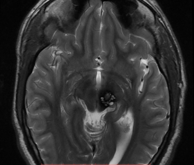
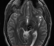
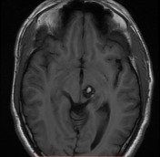
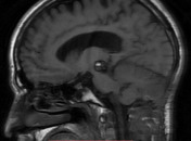
A rounded lesion with peripheral signal drop-out consistent with hemosiderin is seen in left side of midbrain. Centrally the lesion is heterogeneous in signal with regions of high signal on both T1 and T2 weighted images. There is no peripheral edema and no evidence of hydrocephalus.
Features are characteristic of a midbrain cavernous malformation.
Case Discussion
A cerebral cavernous hemangioma is a common type of cerebral vascular malformation. MRI is the modality of choice, demonstrating a characteristic “popcorn” or "berry" appearance with a rim of signal loss due to hemosiderin.


 Unable to process the form. Check for errors and try again.