
Presentation
Work up for dull upper abdominal pain for few months.
Patient Data
Age: 55 years
Gender: Male
From the case:
Cavernous transformation of the portal vein

Show annotations
Download
Info

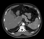
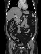
There are multiple serpiginous vascular structures in the porta-hepatis. However, the normal portal vein is not visualized separately.
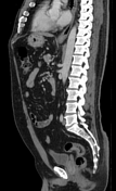
Mild fatty liver and small right Spigelian hernia with small bowel content are noted. No evidence of bowel obstruction.
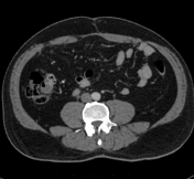
A small right simple renal cyst is visible.
The rest of the major vessels and abdominal viscera are unremarkable.
Case Discussion
Current study shows cavernous transformation of the portal vein due to chronic portal vein thrombosis. This condition is usually associated with portal hypertension.


 Unable to process the form. Check for errors and try again.