Presentation
Heavy smoker complained of chest pain and dyspnea.
Patient Data
Age: 50 years
Gender: Male
From the case:
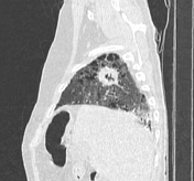
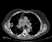
Cavitating pulmonary nodule - lung adenocarcinoma


Download
Info

Irregular thick walled subpleural cavity filled with gas is seen in the right upper lung lobe with adjacent pleural thickening. There is a background of advanced destructive emphysema of both lungs with upper lobe predominance.
Two hepatic focal lesions detected at upper abdominal cuts.
Case Discussion
Transthoracic needle biopsy revealed lung adenocarcinoma. The tumor size was 3 cm in the greatest dimension and hence staged as T1b. The hepatic focal lesions were later diagnosed as hemangiomas.


 Unable to process the form. Check for errors and try again.