Presentation
Fever, shortness of breath.
Patient Data

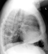
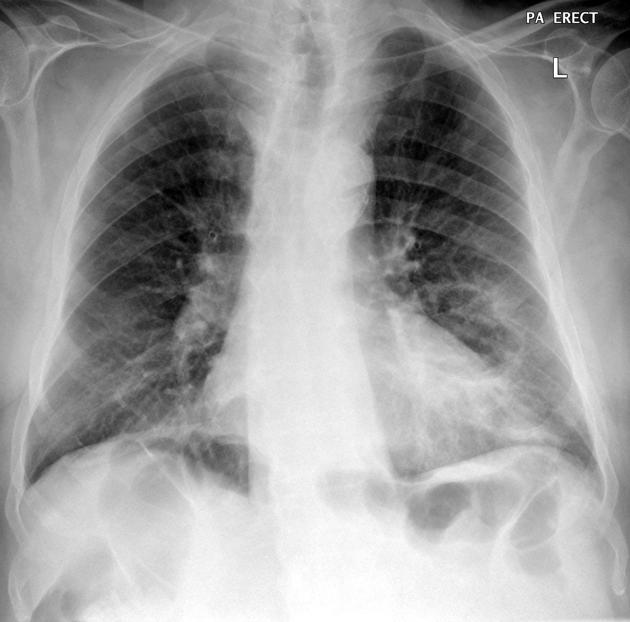
There is a left lower lobe cystic lesion with some irregularity of the wall which has a lobulated contour. It measures 8 x 7 cm. There is minimal adjacent pulmonary opacity. Elsewhere the left lung and pleural space are clear. The right lung and pleural space are clear.
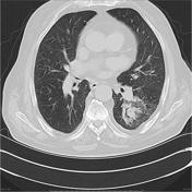

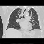

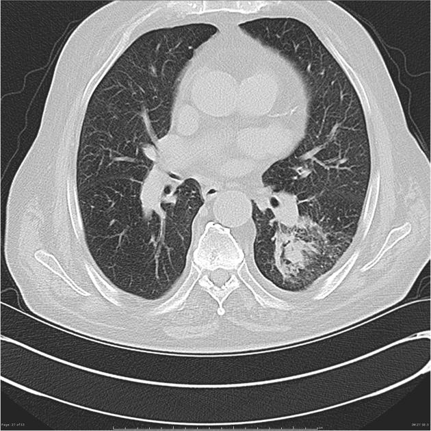
There is a 4.4 x 5.2 x 4.8 cm cavitating lung lesion with nodular irregular borders within the left lower lobe, particularly medially. The wall, where not nodular, is as thin as 1 mm laterally. No fluid level. There is minor associated atelectasis and ground glass peripheral to the lesion along with minor pleural thickening. Adjacent ribs appear normal. No other pulmonary nodules or masses. No lymphadenopathy above the diaphragm. Aortic valve and coronary artery calcification is noted. Anemia.
Conclusion: Solitary LLL cavitating lung lesion.
Case Discussion
Patient proceed to bronchoscopy and cytology demonstrated squamous cell carcinoma.


 Unable to process the form. Check for errors and try again.