
Presentation
The patient presented with right arm and right leg weakness as well as speech and concentration difficulties.
Patient Data


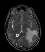
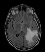
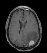

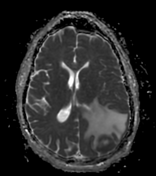
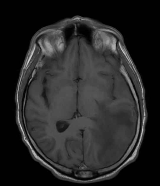
Left inferior parietal lobule intraparenchymal solidly-enhancing mass with evidence of central necrosis. Peripherally, the mass has relatively low T2 signal, high DWI signal, and low ADC signal, suggesting hypercellularity. The mass extends to the cortical margin posteriorly. Some adjacent focal dural thickening is seen. The mass measures 3.2 x 2.7 x 2.9 cm.
Severe surrounding vasogenic edema. Midline shift to the right measuring 6 mm.
Case Discussion
A biopsy of the mass was obtained and pathology revealed diffuse large B-cell lymphoma.
Primary Central Nervous System Lymphoma (PCNSL) is a type of B-cell lymphoma that exclusively affects the brain, spinal cord, and related tissues. The disease's development involves complex immune system interactions and genetic changes. Treatment typically consists of chemotherapy followed by stem cell transplantation or radiation, though treatment options vary based on patient fitness 1.
On MRI, they appear iso-hypointense on T1-weighted images, hypointense on T2-weighted images, and show homogeneous post-gadolinium enhancement. Hemorrhage and calcification are rare but may still occur. Advanced imaging techniques such as Diffusion-Weighted Imaging, Magnetic Resonance Spectroscopy, Magnetic Resonance Perfusion, and Diffusion Tensor Imaging may help in differenting this from other tumors 2.
Case co-author: Alexander Ree, MD, John H. Stroger Hospital of Cook County, Chicago, Illinois, United States


 Unable to process the form. Check for errors and try again.