
Presentation
Increasing headaches.
Patient Data


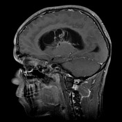
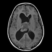

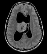
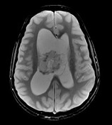
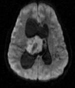
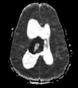

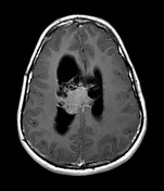
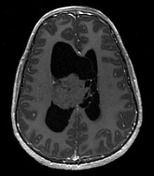
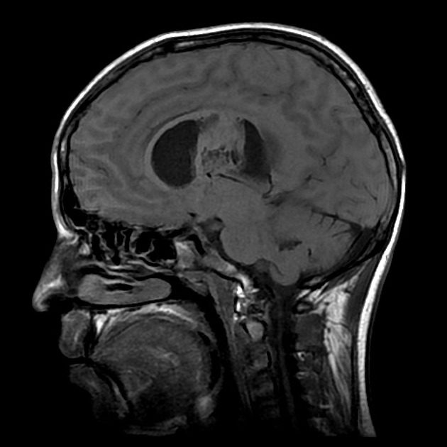
A large mass located in the body of the right lateral ventricle, sending a tongue of tumor to the third ventricle through the right foramen of Monro. It is lobulated with numerous cystic areas. The mass is of similar signal to white matter on T1 and T2 weighted sequences with restricted diffusion and speckled regions of susceptibility induced signal drop out. Following administration of contrast the mass only demonstrates faint heterogeneous enhancement.
The patient went on to have a craniotomy and excision of the mass which confirmed the diagnosis of a central neurocytoma.
Case Discussion
The patient went on to have a craniotomy and excision of the mass which confirmed the diagnosis of a central neurocytoma. This case illustrates typical appearances of a central neurocytoma.


 Unable to process the form. Check for errors and try again.