
Presentation
Headache, drowsiness, and recurrent syncopal attacks.
Patient Data


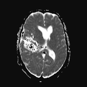
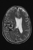
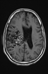
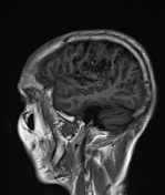

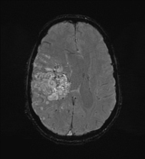
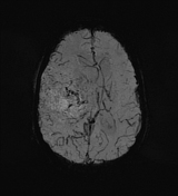
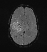
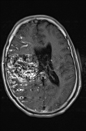
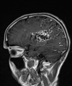
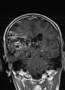
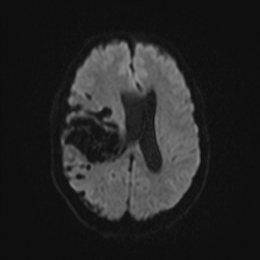
A large bag of worms-like multiple void signals is seen either as convoluted tubular dilated structures or end-on signal voids within the right parietal subcortical and paraventricular region of the right cerebral hemisphere, showing intense enhancement in the post-Gad T1 series. The mentioned complex of tortuous signal void vascular channels measures roughly 6 cm, showing dilated feeding veins from the superior sagittal and straight dural sinuses and to a lesser extent right transverse sinus with feeding arterial supply mainly along the right MCA. The mentioned lesion shows minimal intervening bleeding of high T1 and blooming at SWI, it exerts mass effect with effacement of the right lateral ventricle as well as early sub-falcine herniation.
Case Discussion
Features of MRI findings are consistent with a large cerebral arteriovenous malformation, also known as classic brain AVM, which is a common form of cerebral vascular malformation and is composed of a nidus of tuft vessels through which arteriovenous shunting occurs.


 Unable to process the form. Check for errors and try again.