
Presentation
Headache.
Patient Data
Age: 55 years
Gender: Male
From the case:
Cerebral cavernous venous malformation


Download
Info
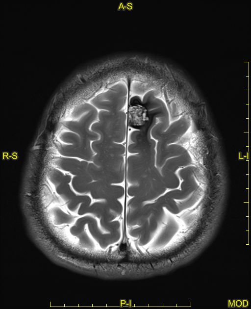
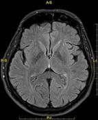
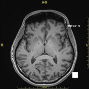
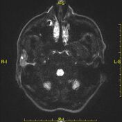
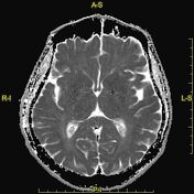
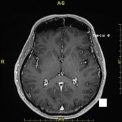
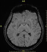
Left frontal lobe well-defined mixed signal intensity lesion, predominantly hyperintense on T2 and FLAIR images with T1 hyperintense foci, giving the popcorn appearance. The lesion is surrounded by complete hemosiderin rim and demonstrates blooming artefact on SWI. No appreciable enhancement could be seen on post contrast study.
No perilesional edema. No significant mass-effect.
No associated developmental venous anomaly (DVA).
The background brain parenchyma shows chronic small vessel ischemic disease.
Case Discussion
Radiological features are mostly in keeping with cerebral cavernous venous malformation (cavernoma).


 Unable to process the form. Check for errors and try again.