
Presentation
Not provided
Patient Data
Age: 25 years
Gender: Female
From the case:
Cerebral cavernous venous malformation


Download
Info
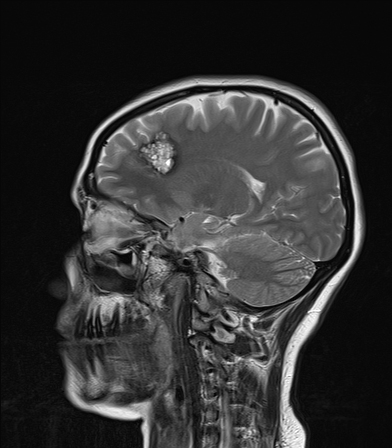
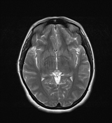
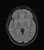
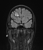
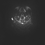
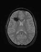
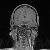
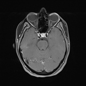
Intra axial well-defined, space-occupying lesion (2.7 x 2.8 x 2.5 cm) in the right frontal lobe that displays typical popcorn appearance on T1 and T2WI with a marginal hypointense rim on T2 due to hemosiderin deposition. The lesion displays susceptibility artifact on T2* GRE. The lesion shows some post-contrast enhancement. A prominent cortical draining vein is seen in its vicinity. No surrounding edema.
The rest of the brain parenchyma is normal.
Case Discussion
The MR imaging features are characteristic of Cerebral cavernous venous malformations, (also called cavernoma) a cerebral vascular malformation.


 Unable to process the form. Check for errors and try again.