
Presentation
Elderly patient with increasing headache.
Patient Data
Age: Elderly
Note: This case has been tagged as "legacy" as it no longer meets image preparation and/or other case publication guidelines.
From the case:
Cerebral metastases - small cell lung cancer

Download
Info
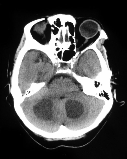
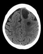
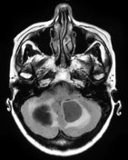
Selected non-contrast CT images demonstrate multiple supra and infratentorial cystic lesions with scant surrounding edema.
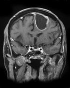
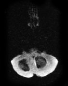
From the case:
Cerebral metastases - small cell lung cancer

Download
Info
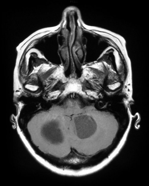
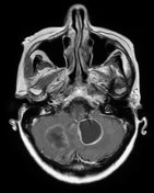
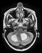
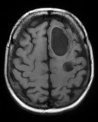
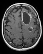
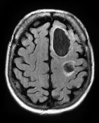
Selected MRI images demonstrate the cystic lesions to have peripheral enhancement, minimal surrounding edema, and no central diffusion restriction. They are most consistent with cystic cerebral metastases.
Case Discussion
This patient had known (histologically confirmed) small cell carcinoma of the lungs. CT and MRI demonstrates multiple cystic cerebral metastases.


 Unable to process the form. Check for errors and try again.