Presentation
Headaches for 5 days, no improvement after medication.
Patient Data


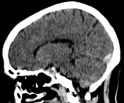

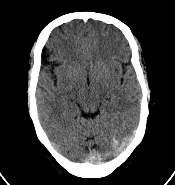
Technique: Volumetric non-contrast axial images have been obtained through the brain.
Findings: There is a heterogeneous hyperdensity in the sinus confluence and along the left transverse venous sinus associated with a slight hypoattenuation along the adjacent occipital lobe parenchyma (cortical edema). There is also a dense strip along the tentorium cerebelli that may correspond to a superficial vein thrombosis; it is hard to define if there are some cortical linear density. The remainder brain parenchyma is unremarkable. Ventricular system is normal for the age group.
Conclusion: Left transverse venous sinus thrombosis with signs of associated adjacent cortical edema.
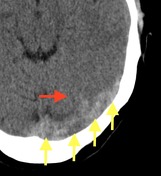
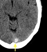
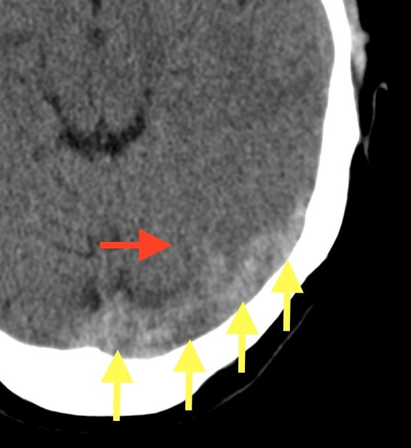
Yellow arrows showing the dense left transverse venous sinus and the sinus confluence. Red arrow spotting the subtle adjacent parenchyma hypoattenuation.
Case Discussion
Unfortunately, we didn't have access to further brain imaging in this patient. The diagnosis is based only on the clinical history and typical CT radiographic features for dural venous sinus thrombosis.
CT and CT venogram can diagnosis venous thrombosis, however, they are less sensitive for assessing venous infarct.


 Unable to process the form. Check for errors and try again.