
Presentation
Upper cervical/base of skull pain.
Patient Data
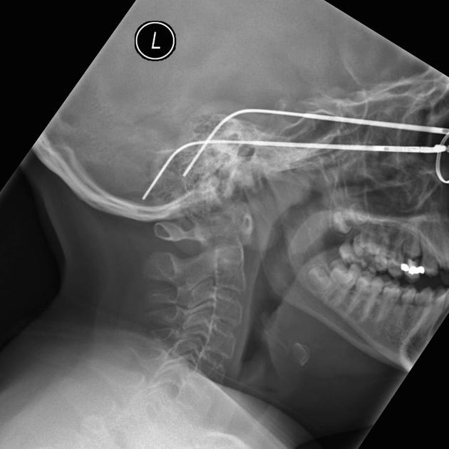
Prominence of the soft tissue in the posterior nasopharynx in this age group most likely represents lymphoid tissue.
The dens and lateral masses of C1 are difficult to discern.


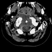
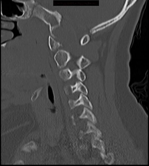
CT reveal extensive bony destruction involving the inferior clivus, occipital condyles, C1 and C2. A large midline soft tissue mass is demonstrated on the soft tissue CT windows. No intratumoral calcifications are identified in this case.
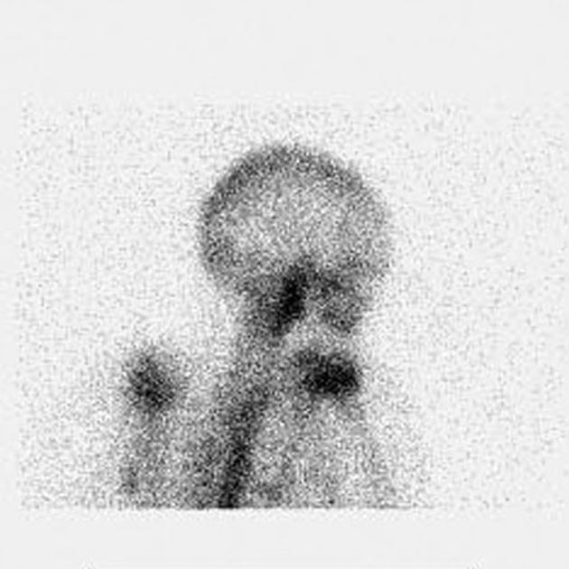
Increased upstake in the region of the upper cervical spine.
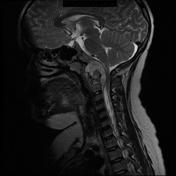
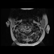
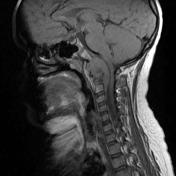
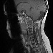
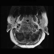
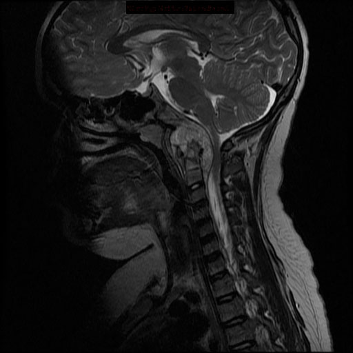
MRI demonstrates an extradural soft tissue mass which involves the basiocciput, anterior arch of C1 and C2 vertebral body. The mass is intermediate to low signal on T1WI, high signal on T2WI and enhances on the post-contrast images. It causes significant mass effect, with compression of the cord at the cervicomedullary junction.
Case Discussion
The mass was biopsied and confirmed the diagnosis of chordoma.
Although chordomas are typically seen in young adults they need to be considered at all age groups, especially when a mass with high T2 signal is encountered, a fairly characteristic appearance.
Case courtesy of Bob Cook, MD. Western Memorial Regional Hospital Corner Brook, Newfoundland.


 Unable to process the form. Check for errors and try again.