
Presentation
Enlarging head size and poor development.
Patient Data
Age: 4 months
Gender: Female


Download
Info

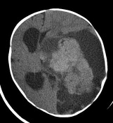
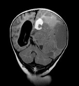
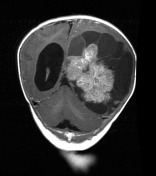
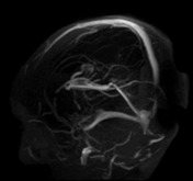
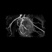
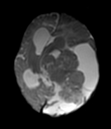
An enormous left sided mass composed of solid enhancing tissue and numerous surrounding cysts is present with enlargement of the skull and marked midline shift.

Download
Info
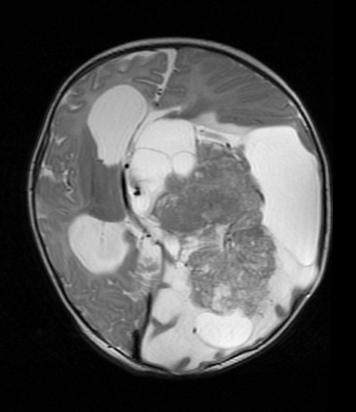
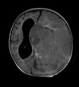
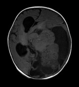
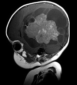
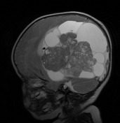
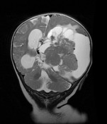
MRI demonstrates a very large heterogeneous mass with solid and cystic components and postcontrast enhancement. No convincing fatty components.
Apologies for the poor contrast / brightness of DWI sequence - the first sequence is DWI followed by B=0
Case Discussion
Typical albeit extreme appearances of a choroid plexus carcinoma.


 Unable to process the form. Check for errors and try again.