
Presentation
Low back pain. History of long-standing recurrent urinary tract infections. No hematuria or fever.
Patient Data


Desiccated L4/L5 intervertebral disc with an annular fissure and posterocentral disc protrusion with narrowing of the bilateral neural foramina. Incidental finding of an exophytic mildly heterogenous lesion (suspicious for a renal malignancy) at the lower pole of the right kidney; this lesion is almost isointense to the renal parenchyma on T2-weighted images. This lesion is also depicted well on the coronal localizer.


Increased parenchymal echogenicity of the right kidney with poor corticomedullary differentiation, particularly at the upper pole. A fine rim of fluid is seen around the upper pole of the right kidney. Please note that the lower pole of the right kidney has not been properly imaged due to excessive bowel gas shadows.
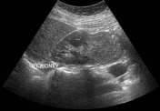


A well-defined exophytic mildly heterogenous solid mass measuring about 4.4 x 4.4 cm is seen at the lower pole of the right kidney. Increased vascularity is seen in it on color doppler ultrasound examination.


A well-defined exophytic mass lesion measuring 4.5 x 4.7 x 6.6 cm is seen along the mid and lower pole of the right kidney. This lesion has an average density of 26, 43 and 85 Hounsfield units on plain, arterial & portovenous phase imaging respectively. Right renal vein is patent and well-opacified. IVC is not well-opacified, likely due to early venous phase imaging. There is no evidence of extension of this lesion into the renal collecting system. No significant locoregional lymphadenopathy or distant metastases is noted.
Multiple surgical clips are seen around the stomach and distal small bowel loops (past history of biliopancreatic surgery). Status post cholecystectomy. An intrauterine contraceptive device (IUCD) is seen in place.
Case Discussion
Procedure: Right radical nephrectomy.
Diagnosis: Chromophobe renal cell carcinoma. Histologic grade: Grade 1. Tumor size: 4 x 5 x 6 cm. Tumor focality: Unifocal. Sarcomatoid/Rhabdoid features: Not identified. Necrosis: Not identified. Tumor extension: Tumor limited to the kidney. Margins: Uninvolved by invasive carcinoma. Lymphovascular invasion: Not identified. Regional lymph nodes: No lymph nodes submitted or found. Pathologic stage: pT1b, pNx.
Additional pathologic findings in non-neoplastic kidney: Tubulointerstitial disease: Chronic pyelonephritis. Immunohistochemical study showed positivity of the tumor with antibody anti-CK7, CD117, CK8 and anti-RCC. Immunostains with the antibody anti-CD10, CK (AE1/AE3), vimentin and CK20 were negative. The special stain with Hale's colloidal iron stain was positive in the tumor cells confirming the diagnosis of chromophobe renal cell carcinoma.
The renal lesion was picked up as an incidental finding on the initial MRI lumbar spine; however, unfortunately, it was not properly communicated with the referring physician (orthopedic surgeon) and not further investigated properly in a timely fashion. Furthermore it was overlooked by the sonographer on first ultrasound as the lower pole of the right kidney was partly obscured by the bowel gases. Ultimately, it was picked up on the follow-up renal ultrasound (16 months after the MRI lumbar spine) and then further investigated with CT urography. The patient was lucky enough that the lesion did not grow rapidly, was localized to the kidney only and was excised completely.


 Unable to process the form. Check for errors and try again.