
Presentation
Palpable mass on left side of abdomen.
Patient Data
Age: 60 years
Gender: Female
From the case:
Clear cell renal cell carcinoma


Download
Info
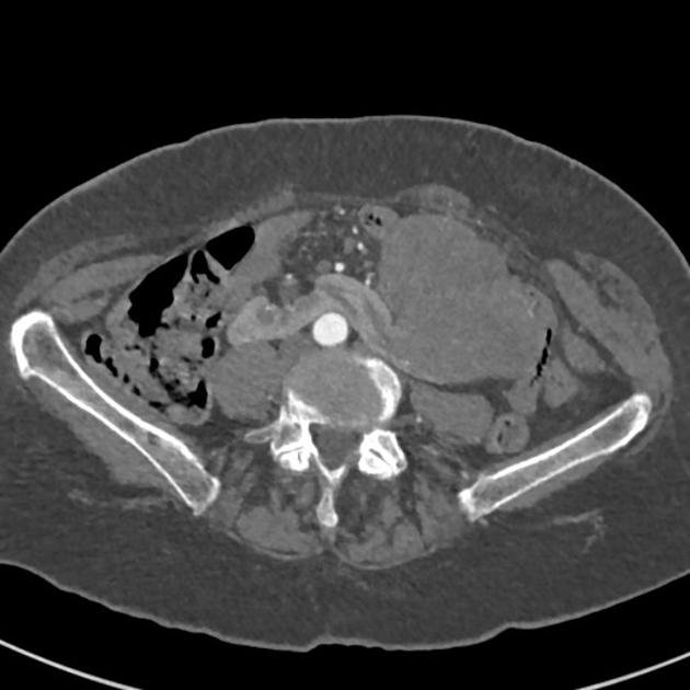
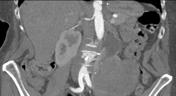
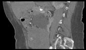
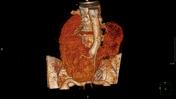
This CT angiogram of the abdomen/pelvis demonstrates a horseshoe kidney with a solid mass originating from the lower pole of the left kidney. Hydronephrosis is also noted on the left kidney.
Case Discussion
This CT angiogram was performed as a pre-operative assessment. The patient subsequently had a left heminephrectomy. Histology showed T3A sarcomatoid renal cell tumor Fuhrman Grade 3.


 Unable to process the form. Check for errors and try again.