
Presentation
Not available.
Patient Data
Note: This case has been tagged as "legacy" as it no longer meets image preparation and/or other case publication guidelines.
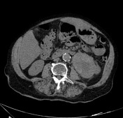
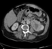
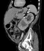

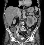

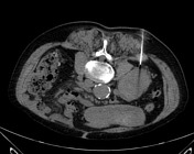
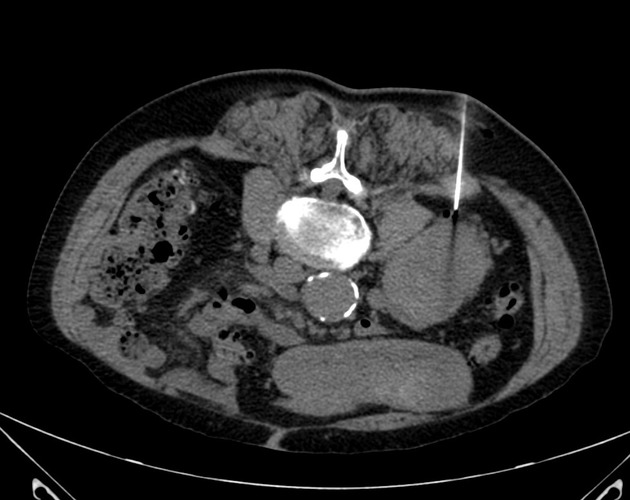
CT scan shows a large mass in the inferior pole of the left kidney, with intense peripheral enhancement and central necrosis.
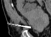
Oncologist team ask for a tumor core-biopsy
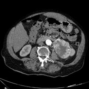
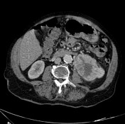
Axial and sagittal view of 18G core-needle biopsy being introduced.
Case Discussion
CT study revealed a large mass in the left kidney with mixed hyperattenuating hypervascular soft tissue components and hypoattenuation areas indicative of necrotic tissue.
A core-needle biopsy was requested by the oncologist. The procedure was made with the patient in ventral decubitus, local anesthesia, and one shot with 18G core-needle, without any complications.
Histopathological study revealed a clear cell renal cell carcinoma.
A surgery approach was proposed.


 Unable to process the form. Check for errors and try again.