Presentation
Respiratory distress and scaphoid abdomen at birth.
Patient Data
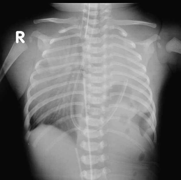
Left hemithorax is opacified with positive mass effect and few lucencies. Additional the contour of left dome of diaphragm not visualized. The nasogastric tube is curled up within the left hemithorax. These features favor congenital diaphragmatic hernia.
Right lung field is hyper-inflated with prominent bronchovascular markings.
The endotracheal tube, umbilical artery and vein catheter are appropriately sited.
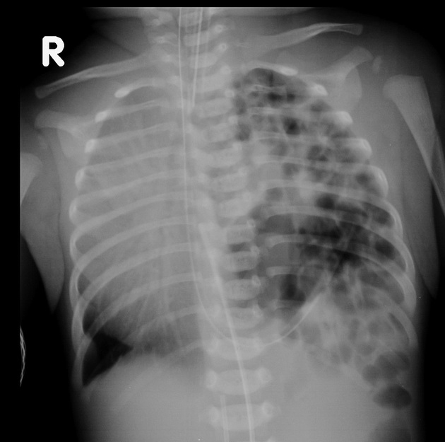
There are multiple cystic lucencies seen within the left hemithorax. The nasogastric tube is curled up within the left hemithorax. The endotracheal tube, umbilical artery and vein catheter are appropriately sited.
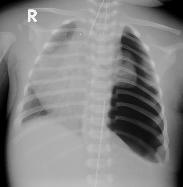
This post-operative film reveals hypo-plastic left lung with surrounding residual air.
The right lung field remains the same.
Again the endotracheal tube and umbilical artery catheter is appropriately sited.
Right upper limb peripherally inserted central catheter insitu.
Intraoperative finds were deficient posterolateral diaphragmatic rim with the following contents - left liver lobe, transverse colon, jejunum, left kidney, spleen and splenenculi.
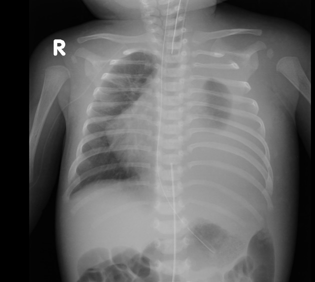
The volume of the left lung remains the same. There is surrounding large volume fluid collection.
Right lung field is hyper-inflated with mild peribronchial thickening in its upper lobe.
Again the endotracheal tube, nasogastric tube and umbilical artery catheter is appropriately sited.
Right upper limb peripherally inserted central catheter insitu.
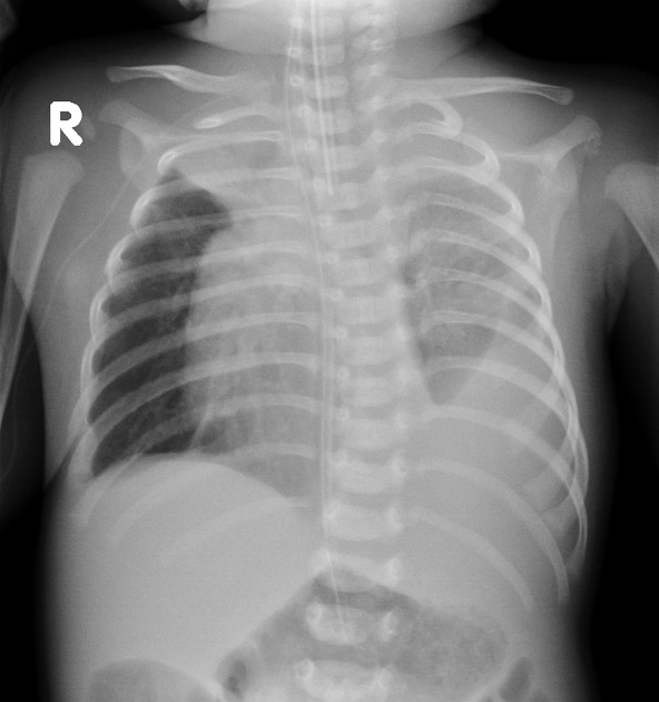
The left lung shows significant expansion. Also the surrounding fluid collection shows mild interval reduction.
Right lung field is hyper-inflated and its upper lobe shows homogenous well defined opacity, suggestive of collapse.
Again the endotracheal tube and nasogastric tube is appropriately sited.
Right upper limb peripherally inserted central catheter insitu.
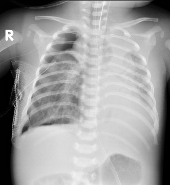
The left lung shows no change in expansion. But surrounding fluid collection appears to be increased.
New finding is right sided pneumothorax. Intercostal drainage tube sited appropriately. Right lung field shows persistent upper lobe collapse. Rest of the lung appears fairly lucent.
The endotracheal tube is seen at T1. Needs re-positioning. Nasogastric tube is appropriately sited.
Right upper limb peripherally inserted central catheter insitu.
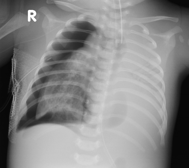
The left lung is partially obscured by surrounding fluid collection, which appears to be comparatively increased.
Right sided pneumothorax has increased. Intercostal drainage tube sited appropriately. Right lung field appears the same as previous radiograph.
The endotracheal tube and nasogastric tube is appropriately sited.
Right upper limb peripherally inserted central catheter insitu.
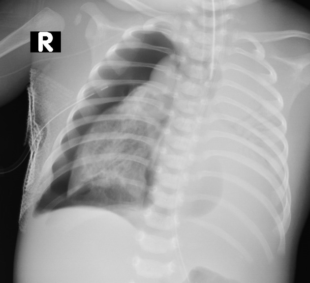
The left lung and surrounding fluid collection remains the same.
Right sided pneumothorax also remains the same. Intercostal drainage tube sited appropriately. Right lung is collapsed with areas of lucency (suggesting a degree of air trapping). **This film was taken minutes before death.
The endotracheal tube and nasogastric tube is appropriately sited.
Right upper limb peripherally inserted central catheter insitu.
Case Discussion
Left sided congenital diaphragmatic hernia (Bochdalek type).
Unfortunately, this infant expired on day 25 of life.


 Unable to process the form. Check for errors and try again.