
Presentation
Admitted with choledocholithiasis. COVID-19 contact. SARS-CoV-2 negative PCR on admission. Three days later cough and fever.
Patient Data


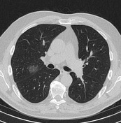
Findings – day 3:
- coronary sclerosis, mild aortic sclerosis
- no significant mediastinal lymphadenopathy
- two small areas of ground-glass opacity (GGO) in the posterior segment of the right upper lobe just above the fissures and in the upper segment of the left lower lobe
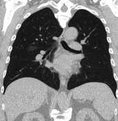
- dilated intrahepatic and extrahepatic bile ducts
Impression:
- Findings are suggestive of COVID-19 pneumonia.
- Biliary obstruction.
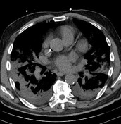
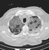
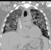

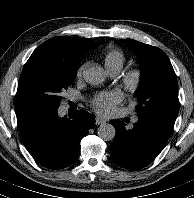
Findings – day 15:
- endotracheal tube and right jugular central catheter in place
- extensive crazy paving pattern with a central and peripheral distribution
- extensive pulmonary consolidations more basal and peripheral
- some mediastinal lymph nodes
- small bilateral pleural effusions
- status post biliary stent with some residual bile duct dilatation
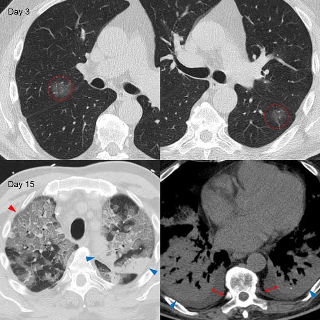
Key findings:
- two small areas of ground-glass opacity on day 3
- extensive crazy-paving pattern (red arrowhead) and consolidations (blue arrowheads) and bilateral pleural effusions (red measurement) on day 15
Case Discussion
A second real-time reverse transcriptase-polymerase chain reaction (PCR) test of the pharyngeal flora just after the first CT was positive for SARS-CoV-2 virus RNA. The patient received ERCP and biliary stent placement because of his biliary obstruction.
Subsequent real-time polymerase chain reaction (PCR) tests on days 12 and 15 were also positive.
This case illustrates a progressive course of COVID-19 pneumonia:
- two very small areas of ground-glass opacity initially
- diffuse involvement of more than 75% of all lobes in both lungs with a typical crazy paving pattern and extensive pulmonary consolidations twelve days after symptom onset
In the second CT, there are also bilateral pleural effusions and some mediastinal lymphadenopathy present, although this is rather atypical for COVID-19 pneumonia.


 Unable to process the form. Check for errors and try again.