
Presentation
8 months of abdominal symptoms with elevated calprotectin suggestive of Crohn disease
Patient Data
Age: 15 years
Gender: Female


Download
Info
MR enterography performed in order to avoid ionizing radiation exposure
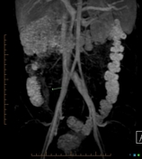
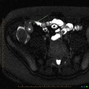
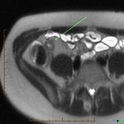
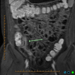
This study shows all the features of Crohn disease, i.e. circumferential ileal wall thickening with abnormal enhancement and inflammation in the adjacent mesentery (comb sign)
Case Discussion
MR enterography has the advantage (over CT enteroclysis and barium follow-through) of no ionizing radiation and can show features of increased disease activity including wall thickening of greater than 4 mm, intramural and mesenteric edema, wall enhancement, fistula formation, and mesenteric vascular engorgement with inflammed mesenteric lymph nodes.


 Unable to process the form. Check for errors and try again.