Presentation
Intermittent fluid from nose.
Patient Data
![](https://prod-images-static.radiopaedia.org/images/19705231/52a0cb68f0c41036acabc5cdc79750_thumb.jpeg)
![This study is a stack](/packs/stack-YQKLCKBI.gif)
![](https://prod-images-static.radiopaedia.org/images/19705317/a5c282f8cb82371e811befa398012b_thumb.jpeg)
![This study is a stack](/packs/stack-YQKLCKBI.gif)
![](https://prod-images-static.radiopaedia.org/images/19705138/ce6060b652f0b23ded41ad7f409cca_thumb.jpeg)
![This study is a stack](/packs/stack-YQKLCKBI.gif)
![](https://prod-images-static.radiopaedia.org/images/19705586/b3ebc5007a3ed92a2786001279ca5a_thumb.jpeg)
![This study is a stack](/packs/stack-YQKLCKBI.gif)
![](https://prod-images-static.radiopaedia.org/images/19706123/044bcc8969c1e4fd62738460167813_thumb.jpeg)
![This study is a stack](/packs/stack-YQKLCKBI.gif)
![](https://prod-images-static.radiopaedia.org/images/19706489/fded23d6e0740463894f39bf116a41_thumb.jpeg)
![This study is a stack](/packs/stack-YQKLCKBI.gif)
![](https://prod-images-static.radiopaedia.org/images/19705344/54acc3f8bcd3bc04af5067a357a980_thumb.jpeg)
![This study is a stack](/packs/stack-YQKLCKBI.gif)
![](https://prod-images-static.radiopaedia.org/images/19705258/b5ad802ae2e8299de867d501a5b7a5_thumb.jpeg)
![This study is a stack](/packs/stack-YQKLCKBI.gif)
![](https://prod-images-static.radiopaedia.org/images/19705114/5304c82f668c01852150e71ebc0698_thumb.jpeg)
![This study is a stack](/packs/stack-YQKLCKBI.gif)
![](https://prod-images-static.radiopaedia.org/images/19705779/02e83a6e000ee21e7cc3386cf1506b_thumb.jpeg)
![This study is a stack](/packs/stack-YQKLCKBI.gif)
![](https://prod-images-static.radiopaedia.org/images/19705290/2ac26232932ac94531944c07a7716c_thumb.jpeg)
![This study is a stack](/packs/stack-YQKLCKBI.gif)
![](https://prod-images-static.radiopaedia.org/images/19705375/06b89674cf8d21378335c9981d8961_thumb.jpeg)
![This study is a stack](/packs/stack-YQKLCKBI.gif)
![](https://prod-images-static.radiopaedia.org/images/19705437/793b69270e1137eb3343e9323b77ae_thumb.jpeg)
![This study is a stack](/packs/stack-YQKLCKBI.gif)
![](https://prod-images-static.radiopaedia.org/images/19705406/3a44ecd6396bb29572db62a1ffce6d_thumb.jpeg)
![This study is a stack](/packs/stack-YQKLCKBI.gif)
![](https://prod-images-static.radiopaedia.org/images/19705186/338ced839fa389533ef60acc2aa0f7_thumb.jpeg)
![This study is a stack](/packs/stack-YQKLCKBI.gif)
![](https://prod-images-static.radiopaedia.org/images/19705231/52a0cb68f0c41036acabc5cdc79750_big_gallery.jpeg)
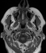
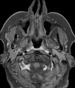
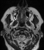
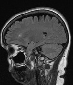
A bony defect is present through the ventromedial aspect of the right middle cranial fossa, through which meninges and distorted brain protrude (meningoencephalocoele), into the lateral aspect of the right side of the sphenoid sinus.
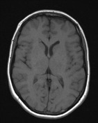
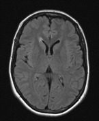
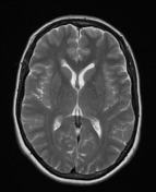
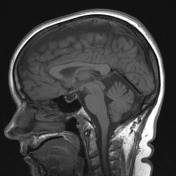
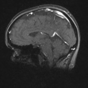
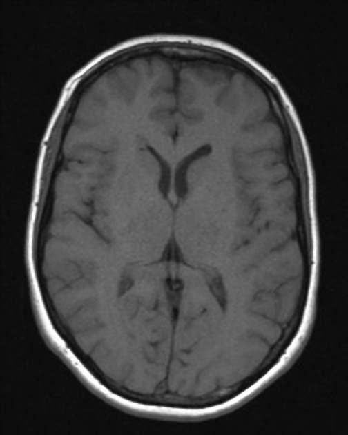
Ancillary findings of intracranial hypertension are also present: empty pituitary fossa, prominent optic nerve sheaths and bilateral distal transverse sinus stenosis.
![](https://prod-images-static.radiopaedia.org/images/19706865/77602afb43ed6680af4be144502d73_thumb.jpeg)
![This study is a stack](/packs/stack-YQKLCKBI.gif)
![](https://prod-images-static.radiopaedia.org/images/19707689/2218d06d5826ef5051f31a2e329485_thumb.jpeg)
![This study is a stack](/packs/stack-YQKLCKBI.gif)
![](https://prod-images-static.radiopaedia.org/images/19706865/77602afb43ed6680af4be144502d73_big_gallery.jpeg)
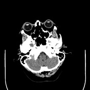
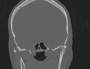
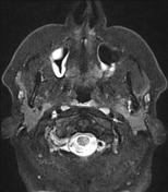
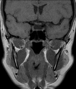
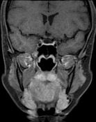
A bony defect is present through the ventromedial aspect of the middle cranial fossa on the right with soft tissue attenuating tissue protruding into the sphenoid sinus.
Case Discussion
This is a classic constellation of findings. A middle aged female with features of intracranial hypertension (pseudotumor cerebri) develops multiple areas of medial sphenoid (middle cranial fossa) scalloping. Eventually one results in a meningocoele or meningoencephalocoele and ruptures into the sphenoid sinus resulting in CSF rhinorrhea.
In many instances the prior elevated intracranial pressures have not been symptomatic or recognized and symptoms become more acute following CSF leak repair (suggesting that the leak was a form of 'self treatment').
The patient went on to have endoscopic repair of the defect at which time CSF was noted to be under unusually high pressures.