
Presentation
Headache and right sided hemiparesis
Patient Data
Age: 35 years
Gender: Female
From the case:
Cystic cerebral metastases

Download
Info

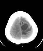
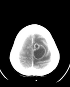
Ring enhancing cystic lesion (37 x 31 mm) in the left high frontal lobe with significant perilesional vasogenic edema causing mass effect in the form of sulcal effacement and subfalcine herniation.
Case Discussion
CT findings are consistent with cystic cerebral metastases in a known case of esophageal carcinoma.
Differential diagnosis for this CT appearance:
MRI (not shown) screening was performed:
- DWI: no restriction in the center of lesion


 Unable to process the form. Check for errors and try again.