
Presentation
Repeated chest infection
Patient Data
Age: 20 years
Gender: Male
From the case:
Cystic fibrosis


Download
Info
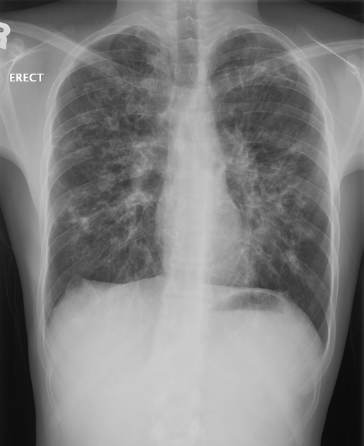
Bilateral predominantly upper lobes bronchiectasis with patchy consolidations and mild hyperinflation.
Case Discussion
Typical findings of cystic fibrosis on plain chest radiographs.


 Unable to process the form. Check for errors and try again.