
Patient Data
Age: 30 months
Gender: Female
From the case:
Developmental dysplasia of the hip


Download
Info
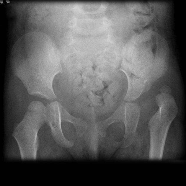
The left hip is dislocated. The femoral head is small and the acetabular rood steeply sloping and poorly formed.
Download
Info

X-ray years after acetabular osteotomy. The femoral epiphysis remains smaller than the right, but it is now well covered by the acetabulum and has developed very well considering the presentation imaging.
Case Discussion
Typical appearances of developmental dysplasia of the hip (DDH) treated with an acetabular osteotomy.
The mainstay of treatment in late-presenting DDH is ensuring that the femoral head is in joint and remains so. This can be achieved with an acetabular osteotomy which increases the volume of the acetabulum and allows the femoral head to sit within the cup of the acetabulum.


 Unable to process the form. Check for errors and try again.