
Presentation
Incidentally discovered, with a history of thyroid cancer.
Patient Data
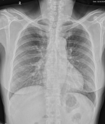
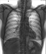

On the chest x-ray, an opacity nodule is noted in the lower left lung, just below the diaphragm, round in shape, with smooth borders and well-defined margins.


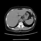
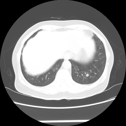
On the CT scan, a soft tissue density nodule was noted in the posterior basal segment (S9) of the left lower lobe, measuring approximately 9 mm, round, with smooth margins and well-defined borders.
Case Discussion
The patient subsequently underwent a PET-CT scan, which revealed a lesion with increased 18-FDG uptake, suggesting a metastatic lesion.
In this specific case, I would like to emphasize the hidden areas on the chest x-ray, which are regions where lesions can easily be overlooked, including:
lung apices, in areas surrounded by the first ribs
hilar zones
retrocardiac areas
lung areas projected beneath the diaphragms


 Unable to process the form. Check for errors and try again.