Presentation
Knee pain. Suspected meniscal tear on bone scintigraphy.
Patient Data


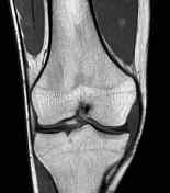

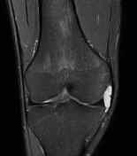

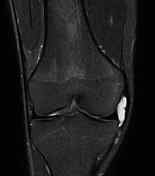

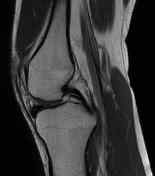

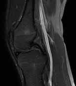

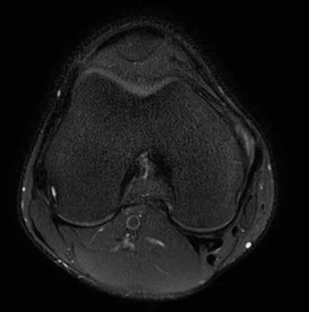
Incomplete discoid medial meniscus. Meniscal cyst along anterior horn. Fine horizontal tear in meniscal body. Medial parameniscal cyst, slightly displacing the MCL. Small cyst in posterior meniscal root (insertional cyst).
Lateral meniscus of normal shape, height and signal.
ACL, PCL, MCL, LCL, quadriceps and patellar tendons of normal thickness, course and signal.
Osseous and joint structures of normal appearance. Muscle layers, fat planes, skin and subcutaneous layer of normal appearance.
Case Discussion
An unusual case of incomplete discoid medial meniscus with a horizontal tear, as the discoid meniscus variant usually affects the lateral meniscus.


 Unable to process the form. Check for errors and try again.