
Presentation
Older gentleman with known duodenal ulcer. 1 day history of upper abdominal pain. Defaulted from surveillance endoscopy and medications.
Patient Data
Age: 75 years
Gender: Male
From the case:
Duodenal perforation


Download
Info
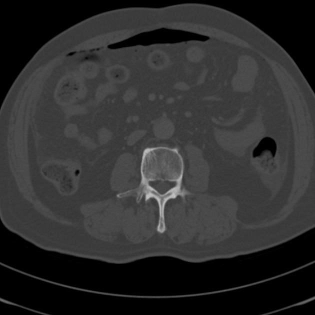
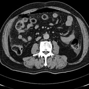
Scattered foci of gas throughout the abdomen with a upper abdominal predominance in keeping with a pneumoperitoneum.
Small volume perihepatic and pelvic free fluid.
Defect in the lateral wall of the 2nd part of the duodenum with a linear gas track at this site suggesting the site of perforation.
Gallstones.
Minor right basal consolidation.
Case Discussion
A laparotomy was performed which identified a 1 cm hole in the second part of the duodenum, consistent with the CT findings.


 Unable to process the form. Check for errors and try again.