
Presentation
Patient with known ulcerative colitis. MRI and CT scan during diagnostic work up.
Patient Data
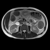


The MRI scan clearly depicts classical signs of ulcerative colitis like the loss of haustral markings (lead pipe sign).
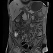
But there is an additional finding. A mass in the fat tissue adjacent to the ascending part of the duodenum. In every sequence the mass shows exactly the same signal intensity and enhancement pattern like the pancreatic tissue. The finding is therefore consistent with ectopic pancreatic tissue.
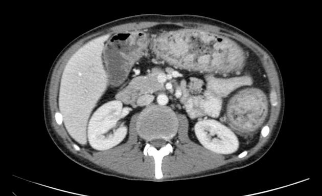
There is a loss of haustral markings (lead pipe sign), the patient has a known chronic ulcerative colitis.
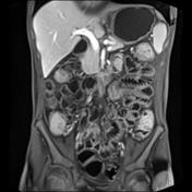
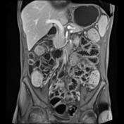
But as in the MRI scan above, the mass in the fat tissue adjacent to the ascending part of the duodenum looks exactly the same as the normal pancreatic tissue. The finding is therefore still consistent with ectopic pancreatic tissue.
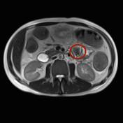
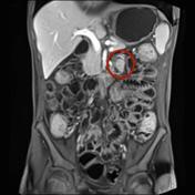


Red circle highlights position of ectopic pancreatic tissue on MR and CT.


 Unable to process the form. Check for errors and try again.