
Presentation
Supratentorial mass.
Patient Data
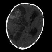

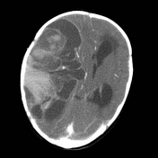
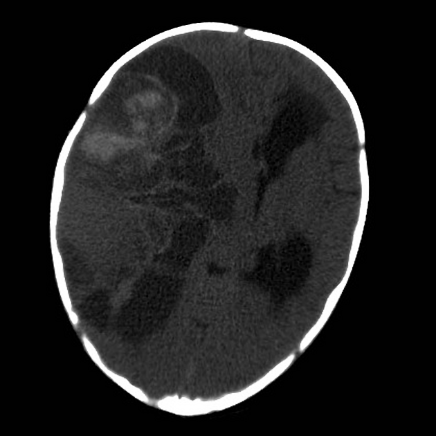
A very large right hemispheric mass, which is markedly heterogeneous in attenuation, with both cystic and hyperdense components is seen; the latter representing hemorrhagic components (see MRI). Following administration of contrast, the mass enhances heterogeneously. Large feeding vessels can be appreciated, including (within the limitations of a non-CTA study) external carotid artery (both superficial temporal artery branches which pass through the thinned parietal bone, and middle meningeal which is markedly enlarged) and middle cerebral artery.
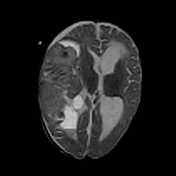
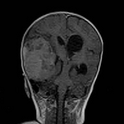
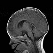

MRI confirms the presence of hemorrhagic components as areas of signal drop out on T2* weighted sequence.
Case Discussion
Given the supratentorial location and large size and heterogeneous appearance, in a young child, the favored diagnosis is a primitive neuroectodermal tumor of the CNS.
Histology was told to confirm the diagnosis of a primitive neuroectodermal tumor of the CNS.
Note: The current (2016) WHO classification of CNS tumors has made substantial changes to tumors previously considered to be PNET, now classified as embryonal tumors with multilayered rosettes (ETMR), along with a number of other entities, in recognition of characteristic amplification of the C19MC region on chromosome 19 (19q13.42).


 Unable to process the form. Check for errors and try again.