
Presentation
Left leg and right flank pain. Later informed about on and off vaginal bleeding.
Patient Data
Age: 65 years
Gender: Female
From the case:
Endometrial adenocarcinoma
Download
Info
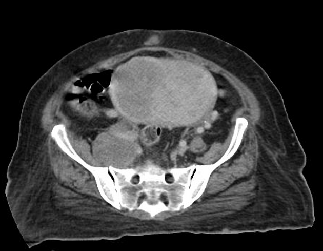
The enhanced CT demonstrates:
- heterogeneously enhancing mass, measuring 12.1 x 7.8 cm, with cystic component, noted in the pelvis
- multiple enlarged para-aortic, aorto-caval, bilateral iliac nodes, largest measuring 5.6 x 4.3 cm
- 3.5 x 3.6 cm omental deposit noted in close proximity to gall bladder
- there is compression of the right distal ureter by the enlarged iliac nodes resulting in moderate-gross right hydroureteronephrosis
- there is compression of bilateral iliac veins by the large iliac nodes (? partially thrombosed)
- with thrombosis of left femoral vein
- right femoral vein does not show any thrombus
Case Discussion
Histopathologically proven case of endometrial adenocarcinoma.


 Unable to process the form. Check for errors and try again.